State Key Laboratory of Ophthalmology, Zhongshan Ophthalmic Center, Sun Yat-sen University, Guangzhou, China.
PLoS One. 2012;7(8):e43373. doi: 10.1371/journal.pone.0043373. Epub 2012 Aug 24.
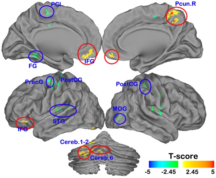
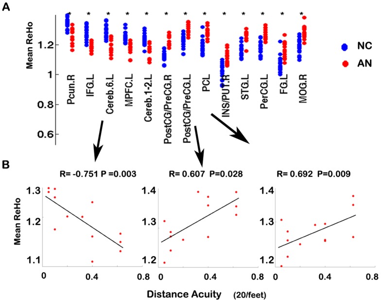
Amblyopia, also known as lazy eye, usually occurs during early childhood and results in poor or blurred vision. Recent neuroimaging studies have found cortical structural/functional abnormalities in amblyopia. However, until now, it was still not known whether the spontaneous activity of the brain changes in amblyopia subjects. In the present study, regional homogeneity (ReHo), a measure of the homogeneity of functional magnetic resonance imaging signals, was used for the first time to investigate changes in resting-state local spontaneous brain activity in individuals with anisometropic amblyopia. Compared with age- and gender-matched subjects with normal vision, the anisometropic amblyopia subjects showed decreased ReHo of spontaneous brain activity in the right precuneus, the left medial prefrontal cortex, the left inferior frontal gyrus, and the left cerebellum, and increased ReHo of spontaneous brain activity was found in the bilateral conjunction area of the postcentral and precentral gyri, the left paracentral lobule, the left superior temporal gyrus, the left fusiform gyrus, the conjunction area of the right insula, putamen and the right middle occipital gyrus. The observed decreases in ReHo may reflect decreased visuo-motor processing ability, and the increases in ReHo in the somatosensory cortices, the motor areas and the auditory area may indicate compensatory plasticity in amblyopia.
弱视,又称懒眼,通常发生在儿童早期,导致视力不佳或模糊。最近的神经影像学研究发现弱视患者存在皮质结构/功能异常。然而,直到现在,仍然不清楚弱视患者的大脑自发活动是否发生变化。在本研究中,首次使用局部一致性(ReHo),一种衡量功能磁共振成像信号均匀性的指标,来研究屈光不正性弱视个体静息状态下局部自发脑活动的变化。与年龄和性别匹配的正常视力受试者相比,屈光不正性弱视受试者右侧楔前叶、左侧内侧前额叶皮质、左侧额下回和左侧小脑的自发脑活动 ReHo 降低,双侧中央后回和中央前回、左侧旁中央小叶、左侧颞上回、左侧梭状回、右侧岛叶、壳核和右侧中枕叶的自发脑活动 ReHo 增加。观察到的 ReHo 降低可能反映了视动处理能力的降低,而感觉皮质、运动区和听觉区的 ReHo 增加可能表明弱视的代偿性可塑性。