Department of Cell Biology, Instituto Nacional de Perinatologia Isidro Espinosa de los Reyes, Mexico City, Mexico.
Reprod Biol Endocrinol. 2012 Sep 3;10:70. doi: 10.1186/1477-7827-10-70.
During intrauterine infection, amniochorionic membranes represent a mechanical and immunological barrier against dissemination of infection. Human beta defensins (HBD)-1, HBD-2, and HBD-3 are key elements of innate immunity that represent the first line of defense against different pathogen microorganisms associated with preterm labor. The aim of this work was to characterize the individual contribution of the amnion (AMN) and choriodecidua (CHD) regions to the secretion of HBD-1, HBD-2 and HBD-3, after stimulation with Candida albicans.
Full-thickness human amniochorionic membranes were obtained after delivery by elective cesarean section from women at 37-40 wk of gestation with no evidence of active labor. The membranes were cultured in a two-compartment experimental model in which the upper compartment is delimited by the amnion and the lower chamber by the choriodecidual membrane. One million of Candida albicans were added to either the AMN or the CHD face or to both and compartmentalized secretion profiles of HBD-1, HBD-2, and HBD-3 were quantified by ELISA. Tissue immunolocalization was performed to detect the presence of HBD-1, -2, -3 in tissue sections stimulated with Candida albicans.
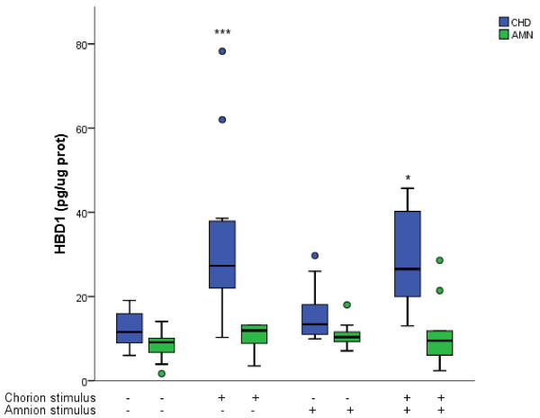
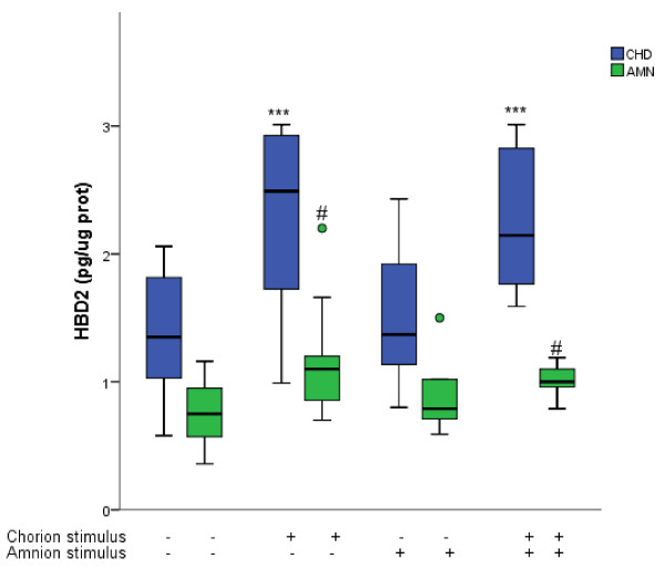
HBD-1 secretion level by the CHD compartment increased 2.6 times (27.30 [20.9-38.25] pg/micrograms protein) when the stimulus with Candida albicans was applied only on this side of the membrane and 2.4 times (26.55 [19.4-42.5] pg/micrograms protein) when applied to both compartments simultaneously. HBD-1 in the amniotic compartment remained without significant changes. HBD-2 secretion level increased significantly in the CHD when the stimulus was applied only to this region (2.49 [1.49-2.95] pg/micrograms protein) and simultaneously to both compartments (2.14 [1.67- 2.91] pg/micrograms protein). When the stimulus was done in the amniotic compartment HBD-2 remained without significant changes in both compartments. HBD-3 remained without significant changes in both compartments regardless of the stimulation modality. Localization of immune-reactive forms of HBD-1, HBD-2, and HBD-3 was carried out by immunohistochemistry confirming the cellular origin of these peptides.
Selective stimulation of amniochorionic membranes with Candida albicans resulted in tissue-specific secretion of HBD-1 and HBD-2, mainly in the CHD, which is the first region to become infected during an ascending infection.
在宫内感染期间,羊膜绒毛膜代表着一种机械和免疫屏障,可防止感染的扩散。人β防御素(HBD)-1、HBD-2 和 HBD-3 是先天免疫的关键因素,是抵御与早产相关的不同病原体微生物的第一道防线。本研究的目的是在与念珠菌刺激后,描述羊膜(AMN)和绒毛膜蜕膜(CHD)区域对 HBD-1、HBD-2 和 HBD-3 分泌的个体贡献。
在 37-40 孕周的择期剖宫产时,从没有活跃分娩迹象的女性中获得完整的羊膜绒毛膜膜。将膜在两室实验模型中培养,其中上室由羊膜限定,下室由绒毛膜蜕膜限定。将 100 万个白色念珠菌加入 AMN 或 CHD 面或两者中,并通过 ELISA 定量测定 HBD-1、HBD-2 和 HBD-3 的分泌谱。通过组织免疫定位检测刺激白色念珠菌后组织切片中 HBD-1、-2、-3 的存在。
当仅在膜的这一侧施加念珠菌刺激时,CHD 隔室的 HBD-1 分泌水平增加了 2.6 倍(27.30 [20.9-38.25] pg/μg 蛋白),当同时施加于两个隔室时增加了 2.4 倍(26.55 [19.4-42.5] pg/μg 蛋白)。在羊膜隔室中,HBD-1 没有明显变化。当刺激仅施加于该区域时,CHD 中的 HBD-2 分泌水平显著增加(2.49 [1.49-2.95] pg/μg 蛋白),同时施加于两个隔室时增加(2.14 [1.67-2.91] pg/μg 蛋白)。当刺激作用于羊膜隔室时,HBD-2 在两个隔室中均无明显变化。无论刺激方式如何,HBD-3 在两个隔室中均无明显变化。通过免疫组织化学进行 HBD-1、HBD-2 和 HBD-3 的免疫反应形式的定位,证实了这些肽的细胞来源。
用白色念珠菌选择性刺激羊膜绒毛膜导致 HBD-1 和 HBD-2 的组织特异性分泌,主要在 CHD 中,CHD 是在上升感染过程中首先感染的区域。