• 文献检索
  • 文档翻译
  • 深度研究
  • 学术资讯
  • Suppr Zotero 插件Zotero 插件
  • 邀请有礼
  • 套餐&价格
  • 历史记录
应用&插件
Suppr Zotero 插件Zotero 插件浏览器插件Mac 客户端Windows 客户端微信小程序
定价
高级版会员购买积分包购买API积分包
服务
文献检索文档翻译深度研究API 文档MCP 服务
关于我们
关于 Suppr公司介绍联系我们用户协议隐私条款
关注我们

Suppr 超能文献

核心技术专利:CN118964589B侵权必究
粤ICP备2023148730 号-1Suppr @ 2026

文献检索

告别复杂PubMed语法,用中文像聊天一样搜索,搜遍4000万医学文献。AI智能推荐,让科研检索更轻松。

立即免费搜索

文件翻译

保留排版,准确专业,支持PDF/Word/PPT等文件格式,支持 12+语言互译。

免费翻译文档

深度研究

AI帮你快速写综述,25分钟生成高质量综述,智能提取关键信息,辅助科研写作。

立即免费体验

使用超薄标本荧光图像的贝叶斯恢复技术实现亚衍射极限下体积空间中蛋白质的定位。

Sub-diffraction limit localization of proteins in volumetric space using Bayesian restoration of fluorescence images from ultrathin specimens.

机构信息

Department of Molecular and Cellular Physiology, Beckman Center, Stanford University, Palo Alto, California, USA.

出版信息

PLoS Comput Biol. 2012;8(8):e1002671. doi: 10.1371/journal.pcbi.1002671. Epub 2012 Aug 30.

DOI:10.1371/journal.pcbi.1002671
PMID:22956902
原文链接:https://pmc.ncbi.nlm.nih.gov/articles/PMC3431294/
Abstract

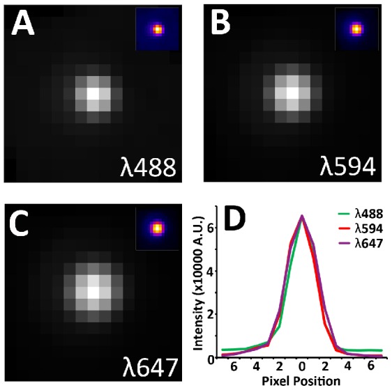
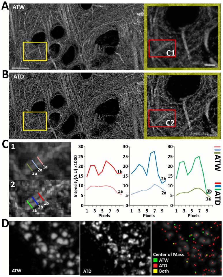
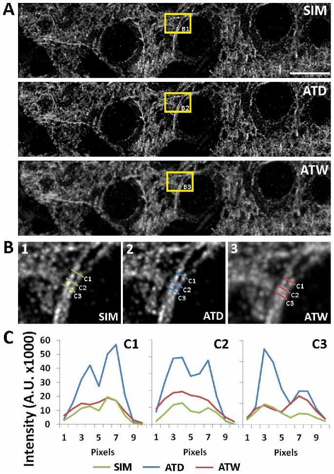
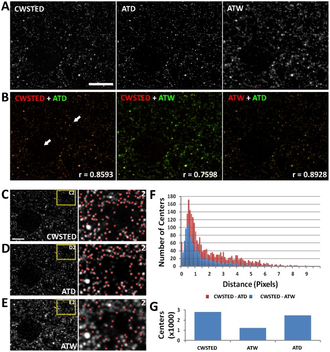
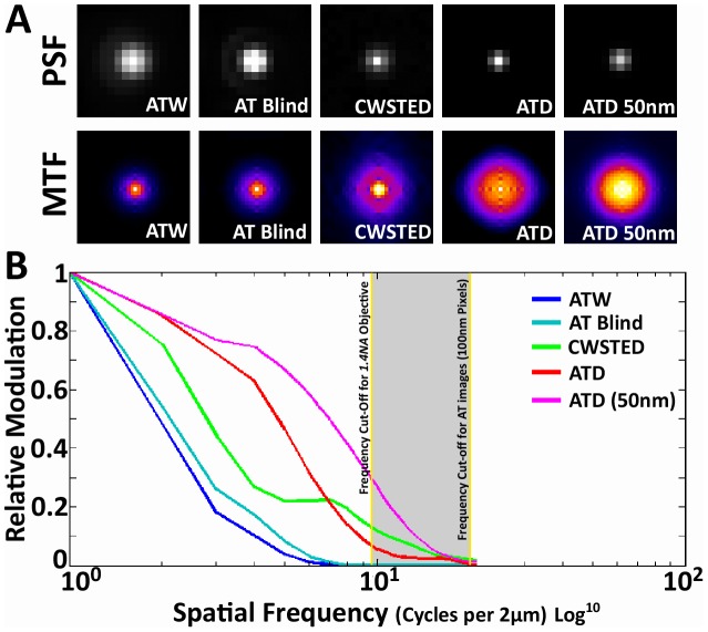
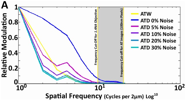
Photon diffraction limits the resolution of conventional light microscopy at the lateral focal plane to 0.61λ/NA (λ = wavelength of light, NA = numerical aperture of the objective) and at the axial plane to 1.4nλ/NA(2) (n = refractive index of the imaging medium, 1.51 for oil immersion), which with visible wavelengths and a 1.4NA oil immersion objective is -220 nm and -600 nm in the lateral plane and axial plane respectively. This volumetric resolution is too large for the proper localization of protein clustering in subcellular structures. Here we combine the newly developed proteomic imaging technique, Array Tomography (AT), with its native 50-100 nm axial resolution achieved by physical sectioning of resin embedded tissue, and a 2D maximum likelihood deconvolution method, based on Bayes' rule, which significantly improves the resolution of protein puncta in the lateral plane to allow accurate and fast computational segmentation and analysis of labeled proteins. The physical sectioning of AT allows tissue specimens to be imaged at the physical optimum of modern high NA plan-apochormatic objectives. This translates to images that have little out of focus light, minimal aberrations and wave-front distortions. Thus, AT is able to provide images with truly invariant point spread functions (PSF), a property critical for accurate deconvolution. We show that AT with deconvolution increases the volumetric analytical fidelity of protein localization by significantly improving the modulation of high spatial frequencies up to and potentially beyond the spatial frequency cut-off of the objective. Moreover, we are able to achieve this improvement with no noticeable introduction of noise or artifacts and arrive at object segmentation and localization accuracies on par with image volumes captured using commercial implementations of super-resolution microscopes.

摘要

光子衍射将传统的明场显微镜在横向焦平面的分辨率限制在 0.61λ/NA(λ=光的波长,NA=物镜的数值孔径),在轴向平面的分辨率限制在 1.4nλ/NA(2)(n=成像介质的折射率,油浸时为 1.51),对于可见波长和 1.4NA 油浸物镜,在横向平面和轴向平面的分辨率分别为-220nm 和-600nm。这种体积分辨率对于亚细胞结构中蛋白质聚簇的正确定位来说太大了。在这里,我们将新开发的蛋白质组学成像技术——阵列断层成像(AT)与它通过对树脂包埋组织进行物理切片获得的 50-100nm 的固有轴向分辨率相结合,以及一种基于贝叶斯规则的 2D 最大似然反卷积方法,该方法显著提高了蛋白质斑点在横向平面上的分辨率,从而能够准确快速地对标记蛋白进行计算分割和分析。AT 的物理切片允许对组织标本在现代高 NA 平面消色差物镜的物理最佳状态下进行成像。这转化为具有很少离焦光、最小像差和波前变形的图像。因此,AT 能够提供具有真正不变的点扩散函数(PSF)的图像,这是准确反卷积的关键属性。我们表明,通过反卷积,AT 增加了蛋白质定位的体积分析保真度,显著提高了高达并可能超过物镜空间频率截止的高空间频率的调制。此外,我们能够在不引入明显噪声或伪影的情况下实现这种改进,并达到与使用商业实施的超分辨率显微镜捕获的图像体积相当的对象分割和定位精度。

https://cdn.ncbi.nlm.nih.gov/pmc/blobs/7344/3431294/d88b19b818fe/pcbi.1002671.g009.jpg
https://cdn.ncbi.nlm.nih.gov/pmc/blobs/7344/3431294/7021d1239331/pcbi.1002671.g001.jpg
https://cdn.ncbi.nlm.nih.gov/pmc/blobs/7344/3431294/26abdd303cc3/pcbi.1002671.g002.jpg
https://cdn.ncbi.nlm.nih.gov/pmc/blobs/7344/3431294/ea95c4cf3146/pcbi.1002671.g003.jpg
https://cdn.ncbi.nlm.nih.gov/pmc/blobs/7344/3431294/dbab4d24bbe4/pcbi.1002671.g004.jpg
https://cdn.ncbi.nlm.nih.gov/pmc/blobs/7344/3431294/ec77d7fb7e00/pcbi.1002671.g005.jpg
https://cdn.ncbi.nlm.nih.gov/pmc/blobs/7344/3431294/c9434c971536/pcbi.1002671.g006.jpg
https://cdn.ncbi.nlm.nih.gov/pmc/blobs/7344/3431294/6d3b1a1667dc/pcbi.1002671.g007.jpg
https://cdn.ncbi.nlm.nih.gov/pmc/blobs/7344/3431294/ced8d5687441/pcbi.1002671.g008.jpg
https://cdn.ncbi.nlm.nih.gov/pmc/blobs/7344/3431294/d88b19b818fe/pcbi.1002671.g009.jpg

相似文献

1
Sub-diffraction limit localization of proteins in volumetric space using Bayesian restoration of fluorescence images from ultrathin specimens.使用超薄标本荧光图像的贝叶斯恢复技术实现亚衍射极限下体积空间中蛋白质的定位。
PLoS Comput Biol. 2012;8(8):e1002671. doi: 10.1371/journal.pcbi.1002671. Epub 2012 Aug 30.
2
High-definition mapping of neural activity using voltage-sensitive dyes.利用电压敏感染料进行神经活动的高清映射。
Methods. 2000 Aug;21(4):349-72. doi: 10.1006/meth.2000.1024.
3
Three dimensional live-cell STED microscopy at increased depth using a water immersion objective.使用水浸物镜在增加深度下进行三维活细胞受激发射损耗显微镜成像。
Rev Sci Instrum. 2018 May;89(5):053701. doi: 10.1063/1.5020249.
4
A Confocal Reflection Super-Resolution Technique to Image Golgi-Cox Stained Neurons.共聚焦反射超分辨率技术对高尔基-考克斯染色神经元进行成像。
J Microsc. 2019 Aug;275(2):115-130. doi: 10.1111/jmi.12821. Epub 2019 Jul 11.
5
Computational resolution in single molecule localization - impact of noise level and emitter density.单分子定位的计算分辨率 - 噪声水平和发射器密度的影响。
Biol Chem. 2023 Feb 14;404(5):427-431. doi: 10.1515/hsz-2022-0301. Print 2023 Apr 25.
6
Shack-Hartmann wave front measurements in cortical tissue for deconvolution of large three-dimensional mosaic transmitted light brightfield micrographs.用于对大型三维镶嵌透射光明场显微照片进行反卷积的皮质组织中的夏克-哈特曼波前测量。
J Microsc. 2009 Feb;233(2):275-89. doi: 10.1111/j.1365-2818.2009.03118.x.
7
Three-Dimensional High-Resolution Digital Inline Hologram Reconstruction with a Volumetric Deconvolution Method.基于体三维反卷积的高分辨率数字全息三维重建。
Sensors (Basel). 2018 Sep 3;18(9):2918. doi: 10.3390/s18092918.
8
Video-rate scanning confocal microscopy and microendoscopy.视频速率扫描共聚焦显微镜和微型内窥镜检查。
J Vis Exp. 2011 Oct 20(56):3252. doi: 10.3791/3252.
9
Mirror-enhanced super-resolution microscopy.镜像增强超分辨率显微镜。
Light Sci Appl. 2016;5(6):e16134-. doi: 10.1038/lsa.2016.134. Epub 2016 Jun 17.
10
Efficient super-resolution volumetric imaging by radial fluctuation Bayesian analysis light-sheet microscopy.径向波动贝叶斯分析光片显微镜实现高效超分辨率体积成像。
J Biophotonics. 2020 Aug;13(8):e201960242. doi: 10.1002/jbio.201960242. Epub 2020 Jun 4.

引用本文的文献

1
Array tomography: trails to discovery.阵列断层扫描:探索之路
Methods Microsc. 2024 Jul 17;1(1):9-17. doi: 10.1515/mim-2024-0001. eCollection 2024 Apr.
2
Chronic Desipramine Reverses Deficits in Cell Activity, Norepinephrine Innervation, and Anxiety-Depression Phenotypes in Fluoxetine-Resistant cF1ko Mice.慢性去甲丙咪嗪逆转氟西汀耐药 cF1ko 小鼠细胞活性、去甲肾上腺素神经支配和焦虑抑郁表型的缺陷。
J Neurosci. 2024 Jan 17;44(3):e1147232023. doi: 10.1523/JNEUROSCI.1147-23.2023.
3
Hyperexcitable arousal circuits drive sleep instability during aging.

本文引用的文献

1
Three-dimensional, tomographic super-resolution fluorescence imaging of serially sectioned thick samples.三维断层超分辨率荧光成像技术在厚样品连续切片中的应用。
PLoS One. 2012;7(5):e38098. doi: 10.1371/journal.pone.0038098. Epub 2012 May 25.
2
Resolution and optical sectioning in the confocal microscope.共聚焦显微镜中的分辨率和光学切片。
J Microsc. 2011 Nov;244(2):113-21. doi: 10.1111/j.1365-2818.2011.03549.x.
3
Single-synapse analysis of a diverse synapse population: proteomic imaging methods and markers.单细胞分析多样性突触群体:蛋白质组学成像方法和标记物。
过度兴奋的唤醒回路导致衰老过程中的睡眠不稳定。
Science. 2022 Feb 25;375(6583):eabh3021. doi: 10.1126/science.abh3021.
4
Fluoxetine-induced recovery of serotonin and norepinephrine projections in a mouse model of post-stroke depression.氟西汀诱导卒中后抑郁小鼠模型中 5-羟色胺和去甲肾上腺素投射的恢复。
Transl Psychiatry. 2020 Sep 30;10(1):334. doi: 10.1038/s41398-020-01008-9.
5
Q&A: Array tomography.常见问题解答:面绘制技术
BMC Biol. 2018 Sep 6;16(1):98. doi: 10.1186/s12915-018-0560-1.
6
Opportunities and challenges for using the zebrafish to study neuronal connectivity as an endpoint of developmental neurotoxicity.利用斑马鱼研究发育神经毒性的神经元连接作为终点的机遇与挑战。
Neurotoxicology. 2018 Jul;67:102-111. doi: 10.1016/j.neuro.2018.04.016. Epub 2018 Apr 25.
7
Probabilistic fluorescence-based synapse detection.基于概率荧光的突触检测。
PLoS Comput Biol. 2017 Apr 17;13(4):e1005493. doi: 10.1371/journal.pcbi.1005493. eCollection 2017 Apr.
8
Sub-synaptic, multiplexed analysis of proteins reveals Fragile X related protein 2 is mislocalized in KO synapses.蛋白质的突触下多重分析表明脆性X相关蛋白2在基因敲除突触中定位错误。
Elife. 2016 Oct 22;5:e20560. doi: 10.7554/eLife.20560.
9
Mapping the connectivity of serotonin transporter immunoreactive axons to excitatory and inhibitory neurochemical synapses in the mouse limbic brain.绘制小鼠边缘脑区中5-羟色胺转运体免疫反应性轴突与兴奋性和抑制性神经化学突触的连接图谱。
Brain Struct Funct. 2017 Apr;222(3):1297-1314. doi: 10.1007/s00429-016-1278-x. Epub 2016 Aug 2.
10
Knowing a synapse when you see one.一眼就能认出突触。
Front Neuroanat. 2015 Jul 28;9:100. doi: 10.3389/fnana.2015.00100. eCollection 2015.
Neuron. 2010 Nov 18;68(4):639-53. doi: 10.1016/j.neuron.2010.09.024.
4
Dependence of 3-D optical transfer functions on the pinhole radius in a fluorescent confocal optical microscope.荧光共焦光学显微镜中三维光学传递函数对针孔半径的依赖性。
Appl Opt. 1990 Jul 10;29(20):3007-11. doi: 10.1364/AO.29.003007.
5
Richardson-Lucy/maximum likelihood image restoration algorithm for fluorescence microscopy: further testing.用于荧光显微镜的理查森-露西/最大似然图像恢复算法:进一步测试
Appl Opt. 1989 Nov 15;28(22):4930-8. doi: 10.1364/AO.28.004930.
6
Three-dimensional resolution doubling in wide-field fluorescence microscopy by structured illumination.通过结构光照实现宽场荧光显微镜三维分辨率翻倍
Biophys J. 2008 Jun;94(12):4957-70. doi: 10.1529/biophysj.107.120345. Epub 2008 Mar 7.
7
STED microscopy with continuous wave beams.连续波光束的受激发射损耗显微镜术
Nat Methods. 2007 Nov;4(11):915-8. doi: 10.1038/nmeth1108. Epub 2007 Oct 21.
8
Array tomography: a new tool for imaging the molecular architecture and ultrastructure of neural circuits.阵列断层扫描:一种用于成像神经回路分子结构和超微结构的新工具。
Neuron. 2007 Jul 5;55(1):25-36. doi: 10.1016/j.neuron.2007.06.014.
9
Nanoscale resolution in GFP-based microscopy.基于绿色荧光蛋白(GFP)的显微镜技术中的纳米级分辨率。
Nat Methods. 2006 Sep;3(9):721-3. doi: 10.1038/nmeth922.
10
STED microscopy reveals that synaptotagmin remains clustered after synaptic vesicle exocytosis.受激发射损耗显微镜显示,突触小泡胞吐后,突触结合蛋白仍聚集在一起。
Nature. 2006 Apr 13;440(7086):935-9. doi: 10.1038/nature04592.