Department of Ophthalmology and Visual Sciences, Havener Eye Institute, The Ohio State University, Columbus, Ohio, United States of America.
PLoS One. 2012;7(9):e44257. doi: 10.1371/journal.pone.0044257. Epub 2012 Sep 6.
Development of retinal detachment models in small animals can be difficult and expensive. Here we create and characterize a novel, cone-rich retinal detachment (RD) model in the chick.
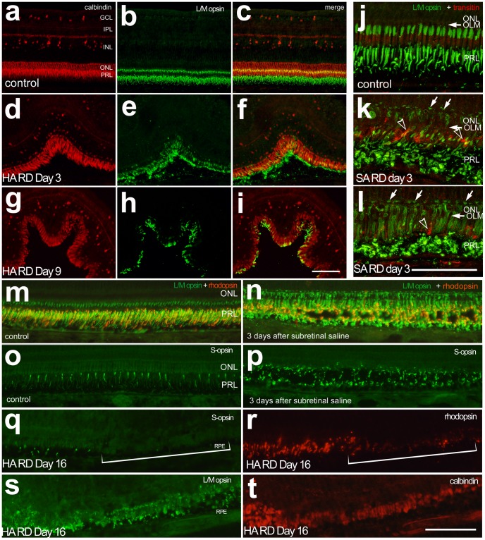
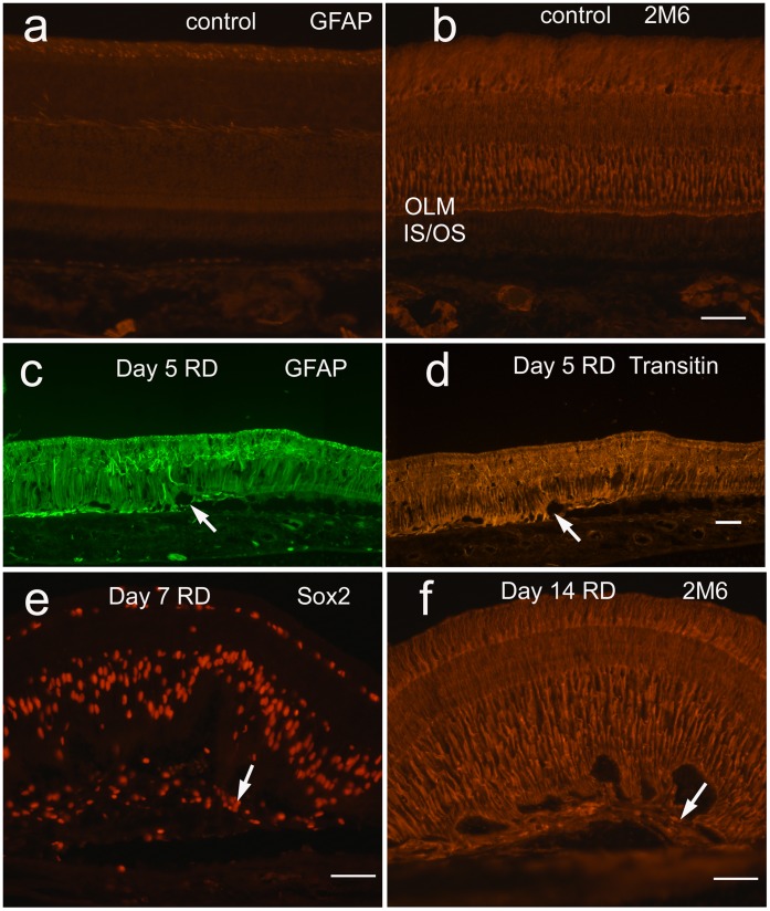
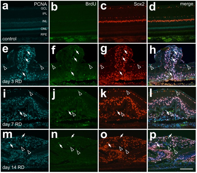
METHODOLOGY/PRINCIPAL FINDINGS: Retinal detachments were created in chicks between postnatal days 7 and 21 by subretinal injections of either saline (SA) or hyaluronic acid (HA). Injections were performed through a dilated pupil with observation via surgical microscope, using the fellow eye as a control. Immunohistochemical analyses were performed at days 1, 3, 7, 10 and 14 after retinal detachment to evaluate the cellular responses of photoreceptors, Müller glia, microglia and nonastrocytic inner retinal glia (NIRG). Cell proliferation was detected with bromodeoxyuridine (BrdU)-incorporation and by the expression of proliferating cell nuclear antigen (PCNA). Cell death was detected with terminal deoxynucleotidyl transferase dUTP nick end labeling (TUNEL). As in mammalian models of RD, there is shortening of photoreceptor outer segments and mis-trafficking of photoreceptor opsins in areas of RD. Photoreceptor cell death was maximal 1 day after RD, but continued until 14 days after RD. Müller glia up-regulated glial fibriliary acidic protein (GFAP), proliferated, showed interkinetic nuclear migration, and migrated to the subretinal space in areas of detachment. Microglia became reactive; they up-regulated CD45, acquired amoeboid morphology, and migrated toward outer retina in areas of RD. Reactive NIRG cells accumulated in detached areas.
CONCLUSIONS/SIGNIFICANCE: Subretinal injections of SA or HA in the chick eye successfully produced retinal detachments and cellular responses similar to those seen in standard mammalian models. Given the relatively large eye size, and considering the low cost, the chick model of RD offers advantages for high-throughput studies.
在小动物中开发视网膜脱离模型可能既困难又昂贵。在这里,我们创建并描述了一种新型的富含锥体的鸡视网膜脱离(RD)模型。
方法/主要发现:通过向出生后 7 至 21 天的小鸡的视网膜下腔注射生理盐水(SA)或透明质酸(HA),在小鸡中创建了视网膜脱离。通过手术显微镜观察,用对侧眼作为对照,在瞳孔扩大时进行注射。在视网膜脱离后第 1、3、7、10 和 14 天进行免疫组织化学分析,以评估光感受器、Müller 胶质细胞、小胶质细胞和非星形胶质内视网膜胶质(NIRG)的细胞反应。通过 BrdU 掺入和增殖细胞核抗原(PCNA)的表达检测细胞增殖。通过末端脱氧核苷酸转移酶 dUTP 缺口末端标记(TUNEL)检测细胞死亡。与哺乳动物 RD 模型一样,在 RD 区域存在光感受器外节缩短和光感受器 opsin 错误运输。RD 后 1 天光感受器细胞死亡达到最大值,但持续至 RD 后 14 天。Müller 胶质细胞上调神经胶质纤维酸性蛋白(GFAP),增殖,表现出核往返迁移,并迁移到脱离区域的视网膜下腔。小胶质细胞变得活跃;它们上调 CD45,获得阿米巴样形态,并向 RD 区域的外视网膜迁移。反应性 NIRG 细胞在分离区域积聚。
结论/意义:在鸡眼中向视网膜下腔注射 SA 或 HA 成功地产生了类似于标准哺乳动物模型中所见的视网膜脱离和细胞反应。鉴于相对较大的眼睛尺寸,并考虑到低成本,鸡 RD 模型为高通量研究提供了优势。