• 文献检索
  • 文档翻译
  • 深度研究
  • 学术资讯
  • Suppr Zotero 插件Zotero 插件
  • 邀请有礼
  • 套餐&价格
  • 历史记录
应用&插件
Suppr Zotero 插件Zotero 插件浏览器插件Mac 客户端Windows 客户端微信小程序
定价
高级版会员购买积分包购买API积分包
服务
文献检索文档翻译深度研究API 文档MCP 服务
关于我们
关于 Suppr公司介绍联系我们用户协议隐私条款
关注我们

Suppr 超能文献

核心技术专利:CN118964589B侵权必究
粤ICP备2023148730 号-1Suppr @ 2026

文献检索

告别复杂PubMed语法,用中文像聊天一样搜索,搜遍4000万医学文献。AI智能推荐,让科研检索更轻松。

立即免费搜索

文件翻译

保留排版,准确专业,支持PDF/Word/PPT等文件格式,支持 12+语言互译。

免费翻译文档

深度研究

AI帮你快速写综述,25分钟生成高质量综述,智能提取关键信息,辅助科研写作。

立即免费体验

功能性磁共振成像证据表明,腹侧前额叶皮层在耳鸣中起作用。

Functional MRI evidence for a role of ventral prefrontal cortex in tinnitus.

机构信息

Department of Neuroscience, Georgetown University Medical Center, 3970 Reservoir Rd. NW, Washington, DC 20007, USA.

出版信息

Brain Res. 2012 Nov 16;1485:22-39. doi: 10.1016/j.brainres.2012.08.052. Epub 2012 Sep 6.

DOI:10.1016/j.brainres.2012.08.052
PMID:22982009
原文链接:https://pmc.ncbi.nlm.nih.gov/articles/PMC3489972/
Abstract

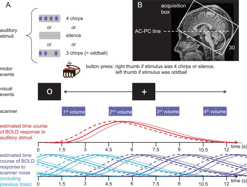
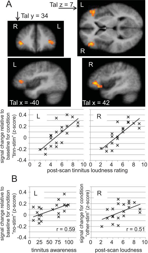
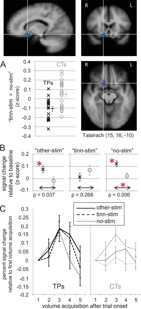
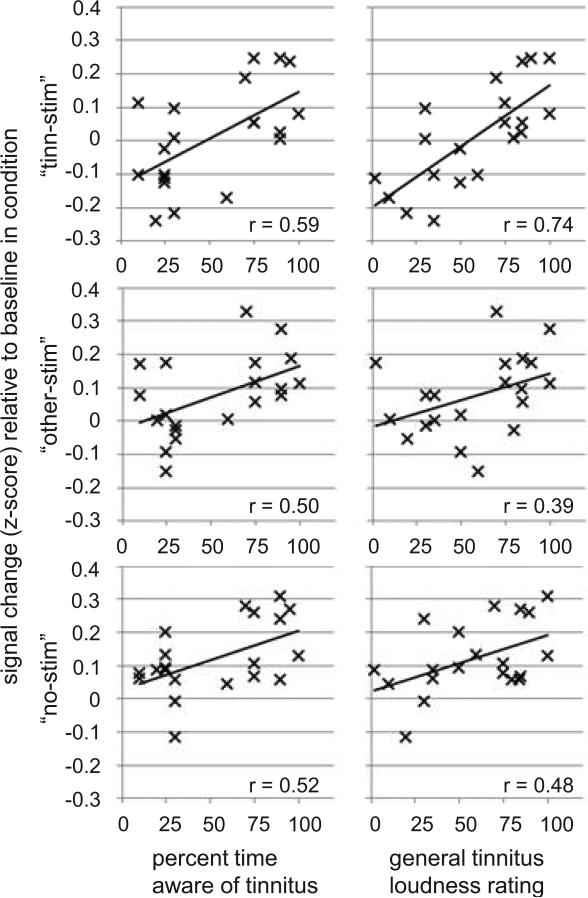
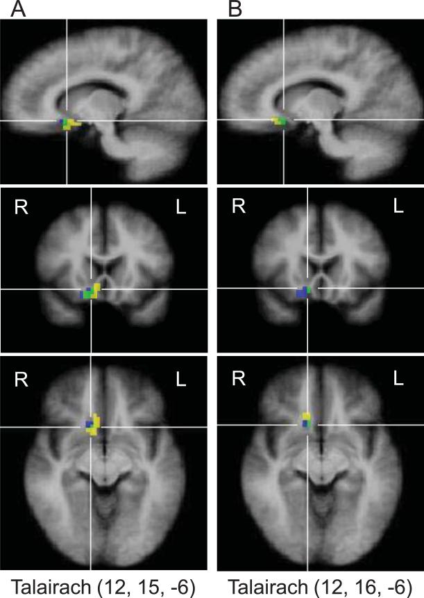
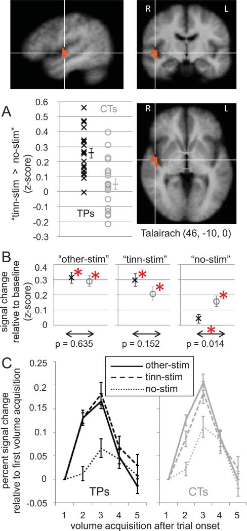
It has long been known that subjective tinnitus, a constant or intermittent phantom sound perceived by 10 to 15% of the adult population, is not a purely auditory phenomenon but is also tied to limbic-related brain regions. Supporting evidence comes from data indicating that stress and emotion can modulate tinnitus, and from brain imaging studies showing functional and anatomical differences in limbic-related brain regions of tinnitus patients and controls. Recent studies from our lab revealed altered blood oxygen level-dependent (BOLD) responses to stimulation at the tinnitus frequency in the ventral striatum (specifically, the nucleus accumbens) and gray-matter reductions (i.e., anatomical changes) in ventromedial prefrontal cortex (vmPFC), of tinnitus patients compared to controls. The present study extended these findings by demonstrating functional differences in vmPFC between 20 tinnitus patients and 20 age-matched controls. Importantly, the observed BOLD response in vmPFC was positively correlated with tinnitus characteristics such as subjective loudness and the percent of time during which the tinnitus was perceived, whereas correlations with tinnitus handicap inventory scores and other variables known to be affected in tinnitus (e.g., depression, anxiety, noise sensitivity, hearing loss) were weaker or absent. This suggests that the observed group differences are indeed related to the strength of the tinnitus percept and not to an affective reaction to tinnitus. The results further corroborate vmPFC as a region of high interest for tinnitus research.This article is part of a Special Issue entitled: Tinnitus Neuroscience.

摘要

长期以来,人们一直知道主观耳鸣,即 10%至 15%的成年人感知到的持续或间歇性幻听,不仅仅是一种纯粹的听觉现象,还与边缘相关的大脑区域有关。有证据表明,压力和情绪可以调节耳鸣,大脑成像研究也显示耳鸣患者和对照组的边缘相关大脑区域在功能和解剖结构上存在差异,这为这一观点提供了支持。我们实验室的最近研究表明,与对照组相比,耳鸣患者的腹侧纹状体(特别是伏隔核)对耳鸣频率的刺激的血氧水平依赖(BOLD)反应发生改变,并且在腹内侧前额叶皮层(vmPFC)中灰质减少(即解剖结构改变)。本研究通过在 20 名耳鸣患者和 20 名年龄匹配的对照组之间的 vmPFC 中显示功能差异,扩展了这些发现。重要的是,vmPFC 中的 BOLD 反应与耳鸣特征(如主观响度和感知耳鸣的时间百分比)呈正相关,而与耳鸣障碍量表评分和其他已知受耳鸣影响的变量(如抑郁、焦虑、噪声敏感性、听力损失)的相关性较弱或不存在。这表明观察到的组间差异确实与耳鸣感知的强度有关,而与对耳鸣的情感反应无关。该结果进一步证实 vmPFC 是耳鸣研究的一个重要区域。本文是特刊“耳鸣神经科学”的一部分。

https://cdn.ncbi.nlm.nih.gov/pmc/blobs/da00/3489972/d4ba2d695694/nihms-405641-f0006.jpg
https://cdn.ncbi.nlm.nih.gov/pmc/blobs/da00/3489972/77f2c1a3abd7/nihms-405641-f0001.jpg
https://cdn.ncbi.nlm.nih.gov/pmc/blobs/da00/3489972/15f47746ea95/nihms-405641-f0002.jpg
https://cdn.ncbi.nlm.nih.gov/pmc/blobs/da00/3489972/37f0b7b86e00/nihms-405641-f0003.jpg
https://cdn.ncbi.nlm.nih.gov/pmc/blobs/da00/3489972/f19c9fb9b2a6/nihms-405641-f0004.jpg
https://cdn.ncbi.nlm.nih.gov/pmc/blobs/da00/3489972/ad97ac72bfc8/nihms-405641-f0005.jpg
https://cdn.ncbi.nlm.nih.gov/pmc/blobs/da00/3489972/d4ba2d695694/nihms-405641-f0006.jpg

相似文献

1
Functional MRI evidence for a role of ventral prefrontal cortex in tinnitus.功能性磁共振成像证据表明,腹侧前额叶皮层在耳鸣中起作用。
Brain Res. 2012 Nov 16;1485:22-39. doi: 10.1016/j.brainres.2012.08.052. Epub 2012 Sep 6.
2
Reduced sound-evoked and resting-state BOLD fMRI connectivity in tinnitus.耳鸣患者听觉诱发和静息状态 fMRI 连接的减少。
Neuroimage Clin. 2018 Aug 31;20:637-649. doi: 10.1016/j.nicl.2018.08.029. eCollection 2018.
3
Altered networks in bothersome tinnitus: a functional connectivity study.恼人耳鸣的改变网络:一项功能连接研究。
BMC Neurosci. 2012 Jan 4;13:3. doi: 10.1186/1471-2202-13-3.
4
Subcallosal brain structure: correlation with hearing threshold at supra-clinical frequencies (>8 kHz), but not with tinnitus.胼胝体下回脑结构:与超临床频率(>8 kHz)的听力阈值相关,而与耳鸣无关。
Hear Res. 2013 Jan;295:79-86. doi: 10.1016/j.heares.2012.03.013. Epub 2012 Apr 7.
5
Diffusion imaging of auditory and auditory-limbic connectivity in tinnitus: preliminary evidence and methodological challenges.耳鸣听神经和听神经边缘连接的弥散成像:初步证据和方法学挑战。
Neural Plast. 2014;2014:145943. doi: 10.1155/2014/145943. Epub 2014 Jun 22.
6
Connectivity graph analysis of the auditory resting state network in tinnitus.耳鸣静息态网络的连通性图谱分析。
Brain Res. 2012 Nov 16;1485:10-21. doi: 10.1016/j.brainres.2012.05.006. Epub 2012 May 10.
7
Ventromedial Prefrontal Cortex Damage Is Associated with Decreased Ventral Striatum Volume and Response to Reward.腹内侧前额叶皮质损伤与腹侧纹状体体积减小及奖励反应降低有关。
J Neurosci. 2016 May 4;36(18):5047-54. doi: 10.1523/JNEUROSCI.4236-15.2016.
8
The Importance of Aging in Gray Matter Changes Within Tinnitus Patients Shown in Cortical Thickness, Surface Area and Volume.衰老在耳鸣患者灰质变化中的重要性:基于皮质厚度、表面积和体积的研究
Brain Topogr. 2016 Nov;29(6):885-896. doi: 10.1007/s10548-016-0511-5. Epub 2016 Aug 10.
9
Dysregulation of limbic and auditory networks in tinnitus.耳鸣的边缘和听觉网络失调。
Neuron. 2011 Jan 13;69(1):33-43. doi: 10.1016/j.neuron.2010.12.002.
10
Feasibility of auditory cortical stimulation for the treatment of tinnitus.听觉皮层刺激治疗耳鸣的可行性。
Otol Neurotol. 2007 Dec;28(8):1005-12. doi: 10.1097/MAO.0b013e318159ebf5.

引用本文的文献

1
Focal tDCS of auditory cortex in chronic tinnitus: A randomized controlled mechanistic trial.听觉皮层聚焦经颅直流电刺激治疗慢性耳鸣的随机对照机制研究
Clin Neurophysiol. 2024 Feb;158:79-91. doi: 10.1016/j.clinph.2023.11.021. Epub 2023 Dec 16.
2
[Progress in neural network mechanism of tinnitus using functional magnetic resonance imaging].[利用功能磁共振成像技术对耳鸣神经网络机制的研究进展]
Lin Chuang Er Bi Yan Hou Tou Jing Wai Ke Za Zhi. 2023 Jul;37(7):582-587. doi: 10.13201/j.issn.2096-7993.2023.07.014.
3
The neuroanatomical hallmarks of chronic tinnitus in comorbidity with pure-tone hearing loss.

本文引用的文献

1
Puzzlingly High Correlations in fMRI Studies of Emotion, Personality, and Social Cognition.令人费解的 fMRI 研究中情绪、个性和社会认知的高度相关性。
Perspect Psychol Sci. 2009 May;4(3):274-90. doi: 10.1111/j.1745-6924.2009.01125.x.
2
Cortico-limbic morphology separates tinnitus from tinnitus distress.皮质-边缘形态将耳鸣与耳鸣困扰区分开来。
Front Syst Neurosci. 2012 Apr 5;6:21. doi: 10.3389/fnsys.2012.00021. eCollection 2012.
3
Phantom percepts: tinnitus and pain as persisting aversive memory networks.幻听感知:耳鸣和疼痛作为持续的厌恶记忆网络。
慢性耳鸣合并纯音听力损失的神经解剖学特征。
Brain Struct Funct. 2023 Jul;228(6):1511-1534. doi: 10.1007/s00429-023-02669-0. Epub 2023 Jun 22.
4
Tinnitus and Influencing Comorbidities.耳鸣及相关共病。
Laryngorhinootologie. 2023 May;102(S 01):S50-S58. doi: 10.1055/a-1950-6149. Epub 2023 May 2.
5
State of the art: non-invasive electrical stimulation for the treatment of chronic tinnitus.前沿技术:用于治疗慢性耳鸣的非侵入性电刺激
Ther Adv Chronic Dis. 2023 Jan 18;14:20406223221148061. doi: 10.1177/20406223221148061. eCollection 2023.
6
Acupuncture decreases amygdala functional connectivity in subjective tinnitus.针刺可降低主观性耳鸣患者杏仁核的功能连接性。
Front Neurol. 2022 Dec 1;13:986805. doi: 10.3389/fneur.2022.986805. eCollection 2022.
7
Invariant structural and functional brain regions associated with tinnitus: A meta-analysis.与耳鸣相关的不变结构和功能脑区:一项荟萃分析。
PLoS One. 2022 Oct 18;17(10):e0276140. doi: 10.1371/journal.pone.0276140. eCollection 2022.
8
Carotenoids improve the development of cerebral cortical networks in formula-fed infant macaques.类胡萝卜素可改善配方奶喂养的猕猴大脑皮质网络的发育。
Sci Rep. 2022 Sep 8;12(1):15220. doi: 10.1038/s41598-022-19279-1.
9
Intrinsic Network Changes in Bilateral Tinnitus Patients with Cognitive Impairment: A Resting-State Functional MRI Study.伴有认知障碍的双侧耳鸣患者的内在网络变化:一项静息态功能磁共振成像研究
Brain Sci. 2022 Aug 8;12(8):1049. doi: 10.3390/brainsci12081049.
10
Tinnitus and the Triple Network Model: A Perspective.耳鸣与三重网络模型:一种观点
Clin Exp Otorhinolaryngol. 2022 Aug;15(3):205-212. doi: 10.21053/ceo.2022.00815. Epub 2022 Jul 11.
Proc Natl Acad Sci U S A. 2011 May 17;108(20):8075-80. doi: 10.1073/pnas.1018466108. Epub 2011 Apr 18.
4
Dysregulation of limbic and auditory networks in tinnitus.耳鸣的边缘和听觉网络失调。
Neuron. 2011 Jan 13;69(1):33-43. doi: 10.1016/j.neuron.2010.12.002.
5
Ringing ears: the neuroscience of tinnitus.耳鸣:神经科学视角下的耳鸣
J Neurosci. 2010 Nov 10;30(45):14972-9. doi: 10.1523/JNEUROSCI.4028-10.2010.
6
Tinnitus, diminished sound-level tolerance, and elevated auditory activity in humans with clinically normal hearing sensitivity.耳鸣、听阈降低和听觉活动增强在临床听力正常的人群中。
J Neurophysiol. 2010 Dec;104(6):3361-70. doi: 10.1152/jn.00226.2010. Epub 2010 Sep 29.
7
Tuning out the noise: limbic-auditory interactions in tinnitus.调谐出噪声:耳鸣的边缘听觉相互作用。
Neuron. 2010 Jun 24;66(6):819-26. doi: 10.1016/j.neuron.2010.04.032.
8
Tinnitus modulation by deep brain stimulation in locus of caudate neurons (area LC).通过深部脑刺激调节尾状核神经元(LC 区)的耳鸣。
Neuroscience. 2010 Sep 15;169(4):1768-78. doi: 10.1016/j.neuroscience.2010.06.007. Epub 2010 Jun 10.
9
Mapping cortical hubs in tinnitus.耳鸣皮质中枢连接图谱。
BMC Biol. 2009 Nov 23;7:80. doi: 10.1186/1741-7007-7-80.
10
Hyperactivity in the auditory midbrain after acoustic trauma: dependence on cochlear activity.听神经损伤后中脑听觉过度兴奋:耳蜗活动依赖性。
Neuroscience. 2009 Dec 1;164(2):733-46. doi: 10.1016/j.neuroscience.2009.08.036. Epub 2009 Aug 20.