Department of Neuroscience, Georgetown University Medical Center, Washington DC, USA.
Front Syst Neurosci. 2012 Apr 5;6:21. doi: 10.3389/fnsys.2012.00021. eCollection 2012.
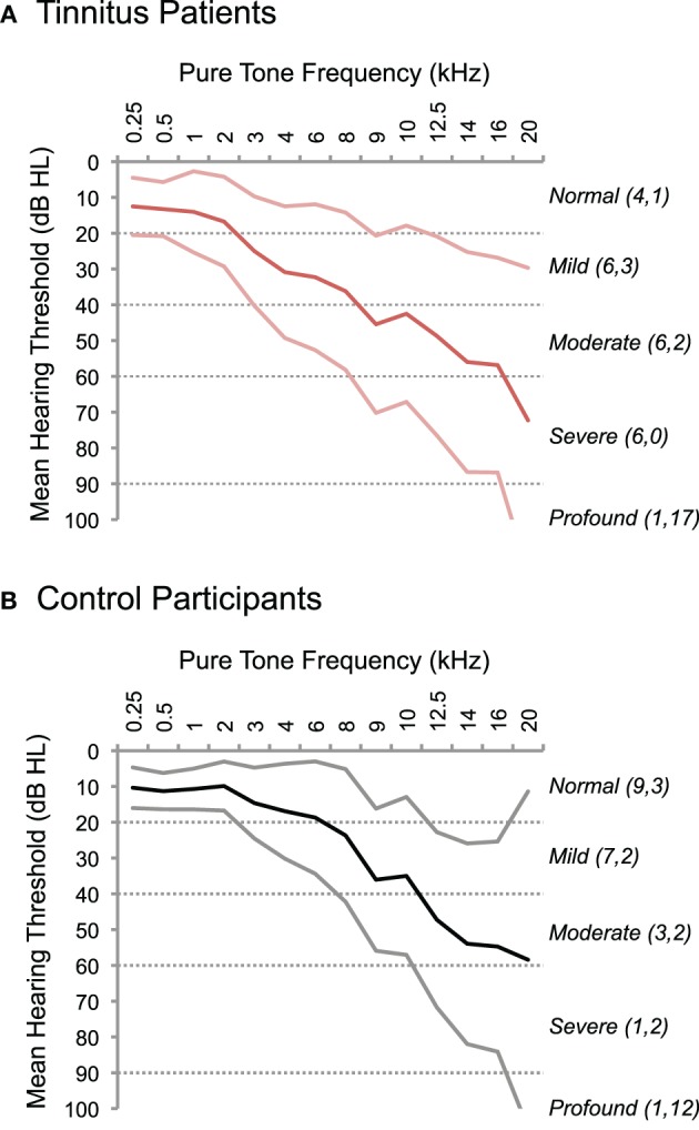
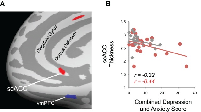
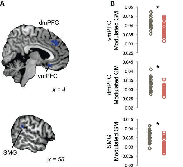
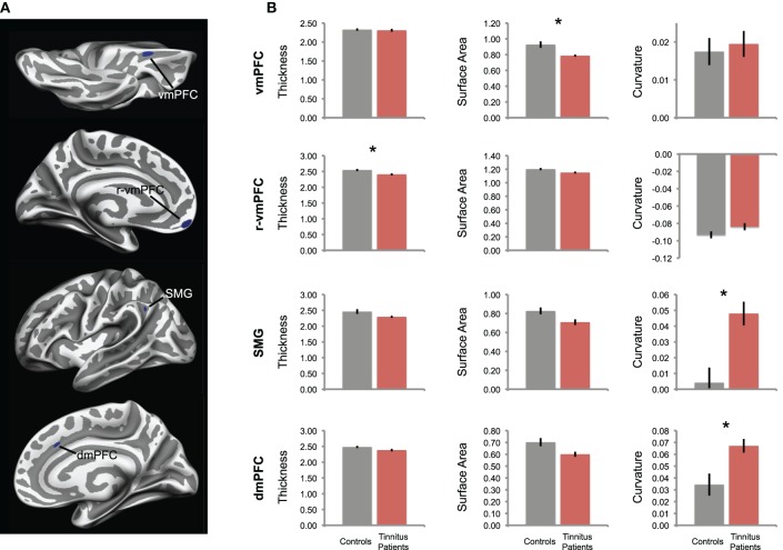
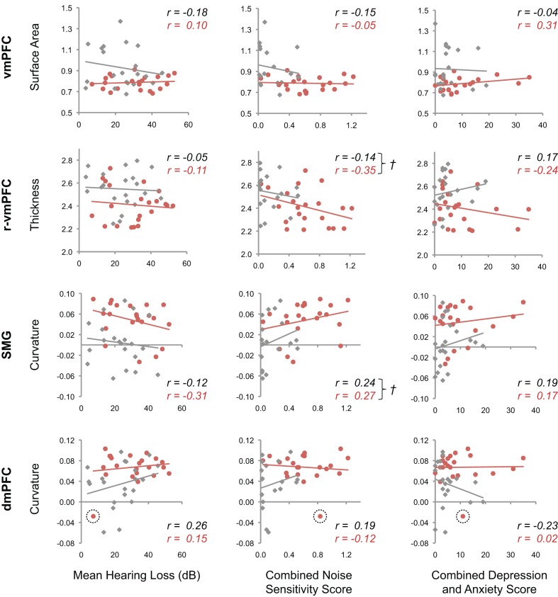
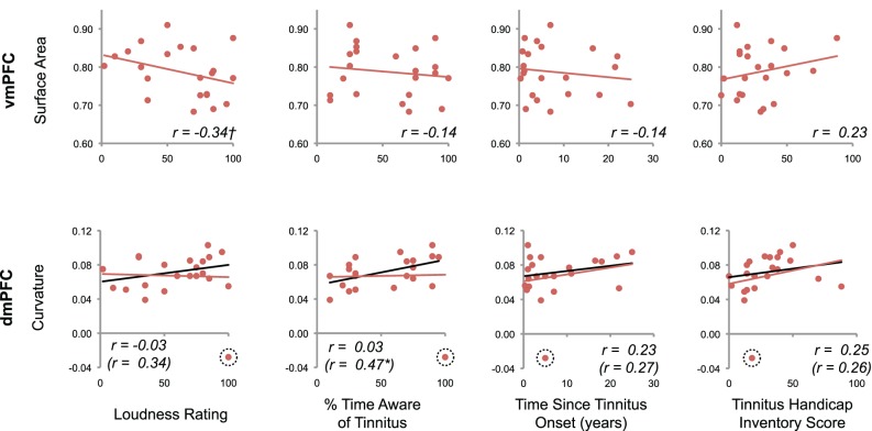
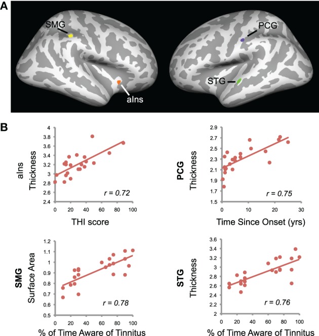
Tinnitus is a common auditory disorder characterized by a chronic ringing or buzzing "in the ear."Despite the auditory-perceptual nature of this disorder, a growing number of studies have reported neuroanatomical differences in tinnitus patients outside the auditory-perceptual system. Some have used this evidence to characterize chronic tinnitus as dysregulation of the auditory system, either resulting from inefficient inhibitory control or through the formation of aversive associations with tinnitus. It remains unclear, however, whether these "non-auditory" anatomical markers of tinnitus are related to the tinnitus signal itself, or merely to negative emotional reactions to tinnitus (i.e., tinnitus distress). In the current study, we used anatomical MRI to identify neural markers of tinnitus, and measured their relationship to a variety of tinnitus characteristics and other factors often linked to tinnitus, such as hearing loss, depression, anxiety, and noise sensitivity. In a new cohort of participants, we confirmed that people with chronic tinnitus exhibit reduced gray matter in ventromedial prefrontal cortex (vmPFC) compared to controls matched for age and hearing loss. This effect was driven by reduced cortical surface area, and was not related to tinnitus distress, symptoms of depression or anxiety, noise sensitivity, or other factors. Instead, tinnitus distress was positively correlated with cortical thickness in the anterior insula in tinnitus patients, while symptoms of anxiety and depression were negatively correlated with cortical thickness in subcallosal anterior cingulate cortex (scACC) across all groups. Tinnitus patients also exhibited increased gyrification of dorsomedial prefrontal cortex (dmPFC), which was more severe in those patients with constant (vs. intermittent) tinnitus awareness. Our data suggest that the neural systems associated with chronic tinnitus are different from those involved in aversive or distressed reactions to tinnitus.
耳鸣是一种常见的听觉障碍,其特征为慢性的“耳内”鸣响或嗡嗡声。尽管这种障碍具有听觉知觉性质,但越来越多的研究报告称,耳鸣患者的听觉知觉系统之外存在神经解剖差异。一些研究人员利用这些证据将慢性耳鸣描述为听觉系统的失调,其原因可能是抑制控制效率低下,也可能是与耳鸣形成厌恶关联。然而,目前尚不清楚这些耳鸣的“非听觉”解剖学标志物是否与耳鸣信号本身有关,或者仅仅与对耳鸣的负面情绪反应(即耳鸣困扰)有关。在当前的研究中,我们使用解剖 MRI 来确定耳鸣的神经标志物,并测量它们与各种耳鸣特征以及其他通常与耳鸣相关的因素(例如听力损失、抑郁、焦虑和噪声敏感性)之间的关系。在一个新的参与者队列中,我们证实与年龄和听力损失相匹配的对照组相比,慢性耳鸣患者的腹内侧前额叶皮质(vmPFC)灰质减少。这种效应是由皮质表面积减少驱动的,与耳鸣困扰、抑郁或焦虑症状、噪声敏感性或其他因素无关。相反,耳鸣困扰与耳鸣患者前脑岛的皮质厚度呈正相关,而焦虑和抑郁症状与所有组的扣带前回皮质下(scACC)的皮质厚度呈负相关。耳鸣患者还表现出背内侧前额叶皮质(dmPFC)的脑回增多,在那些持续(而非间歇性)耳鸣意识的患者中更为严重。我们的数据表明,与慢性耳鸣相关的神经系统与对耳鸣的厌恶或困扰反应所涉及的系统不同。