• 文献检索
  • 文档翻译
  • 深度研究
  • 学术资讯
  • Suppr Zotero 插件Zotero 插件
  • 邀请有礼
  • 套餐&价格
  • 历史记录
应用&插件
Suppr Zotero 插件Zotero 插件浏览器插件Mac 客户端Windows 客户端微信小程序
定价
高级版会员购买积分包购买API积分包
服务
文献检索文档翻译深度研究API 文档MCP 服务
关于我们
关于 Suppr公司介绍联系我们用户协议隐私条款
关注我们

Suppr 超能文献

核心技术专利:CN118964589B侵权必究
粤ICP备2023148730 号-1Suppr @ 2026

文献检索

告别复杂PubMed语法,用中文像聊天一样搜索,搜遍4000万医学文献。AI智能推荐,让科研检索更轻松。

立即免费搜索

文件翻译

保留排版,准确专业,支持PDF/Word/PPT等文件格式,支持 12+语言互译。

免费翻译文档

深度研究

AI帮你快速写综述,25分钟生成高质量综述,智能提取关键信息,辅助科研写作。

立即免费体验

利用沟道衬度机制对极薄层进行氦离子显微镜成像。

Imaging ultra thin layers with helium ion microscopy: Utilizing the channeling contrast mechanism.

机构信息

Physics of Interfaces and Nanomaterials, MESA+ Institute for Nanotechnology, University of Twente, PO Box 217, 7500AE Enschede, The Netherlands.

出版信息

Beilstein J Nanotechnol. 2012;3:507-12. doi: 10.3762/bjnano.3.58. Epub 2012 Jul 12.

DOI:10.3762/bjnano.3.58
PMID:23019545
原文链接:https://pmc.ncbi.nlm.nih.gov/articles/PMC3458595/
Abstract

BACKGROUND

Helium ion microscopy is a new high-performance alternative to classical scanning electron microscopy. It provides superior resolution and high surface sensitivity by using secondary electrons.

RESULTS

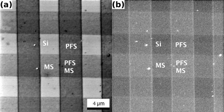
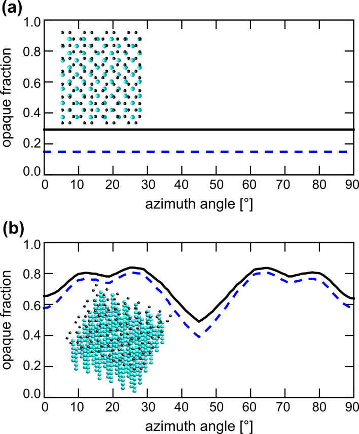
We report on a new contrast mechanism that extends the high surface sensitivity that is usually achieved in secondary electron images, to backscattered helium images. We demonstrate how thin organic and inorganic layers as well as self-assembled monolayers can be visualized on heavier element substrates by changes in the backscatter yield. Thin layers of light elements on heavy substrates should have a negligible direct influence on backscatter yields. However, using simple geometric calculations of the opaque crystal fraction, the contrast that is observed in the images can be interpreted in terms of changes in the channeling probability.

CONCLUSION

The suppression of ion channeling into crystalline matter by adsorbed thin films provides a new contrast mechanism for HIM. This dechanneling contrast is particularly well suited for the visualization of ultrathin layers of light elements on heavier substrates. Our results also highlight the importance of proper vacuum conditions for channeling-based experimental methods.

摘要

背景

氦离子显微镜是一种新型的高性能扫描电子显微镜替代品。它通过使用二次电子提供更高的分辨率和高表面灵敏度。

结果

我们报告了一种新的对比机制,该机制将通常在二次电子图像中实现的高表面灵敏度扩展到背散射氦图像。我们展示了如何通过背散射产额的变化,在较重元素衬底上可视化薄的有机和无机层以及自组装单层。重衬底上的轻元素薄层应该对背散射产额没有直接影响。然而,使用不透明晶体分数的简单几何计算,可以根据沟道概率的变化来解释在图像中观察到的对比度。

结论

吸附的薄膜对离子进入晶体的抑制提供了 HIM 的一种新的对比机制。这种去沟道对比度特别适合于在较重衬底上可视化超薄的轻元素层。我们的结果还强调了适当的真空条件对于基于沟道的实验方法的重要性。

https://cdn.ncbi.nlm.nih.gov/pmc/blobs/bfa2/3458595/817c295295c2/Beilstein_J_Nanotechnol-03-507-g005.jpg
https://cdn.ncbi.nlm.nih.gov/pmc/blobs/bfa2/3458595/00641a3df670/Beilstein_J_Nanotechnol-03-507-g002.jpg
https://cdn.ncbi.nlm.nih.gov/pmc/blobs/bfa2/3458595/640273d105cc/Beilstein_J_Nanotechnol-03-507-g003.jpg
https://cdn.ncbi.nlm.nih.gov/pmc/blobs/bfa2/3458595/53f5e1faed77/Beilstein_J_Nanotechnol-03-507-g004.jpg
https://cdn.ncbi.nlm.nih.gov/pmc/blobs/bfa2/3458595/817c295295c2/Beilstein_J_Nanotechnol-03-507-g005.jpg

相似文献

1
Imaging ultra thin layers with helium ion microscopy: Utilizing the channeling contrast mechanism.利用沟道衬度机制对极薄层进行氦离子显微镜成像。
Beilstein J Nanotechnol. 2012;3:507-12. doi: 10.3762/bjnano.3.58. Epub 2012 Jul 12.
2
Channeling in helium ion microscopy: Mapping of crystal orientation.氦离子显微镜中的束流导向:晶体取向测绘。
Beilstein J Nanotechnol. 2012;3:501-6. doi: 10.3762/bjnano.3.57. Epub 2012 Jul 10.
3
The interaction of a nanoscale coherent helium-ion probe with a crystal.纳米级相干氦离子探针与晶体的相互作用。
Ultramicroscopy. 2013 Nov;134:18-22. doi: 10.1016/j.ultramic.2013.06.019. Epub 2013 Jul 3.
4
Visualization of steps and surface reconstructions in Helium Ion Microscopy with atomic precision.在氦离子显微镜中以原子精度实现步骤可视化和表面重建。
Ultramicroscopy. 2016 Mar;162:17-24. doi: 10.1016/j.ultramic.2015.11.009. Epub 2015 Dec 9.
5
Imaging of carbon nanomembranes with helium ion microscopy.氦离子显微镜对碳纳米膜的成像
Beilstein J Nanotechnol. 2015 Aug 12;6:1712-20. doi: 10.3762/bjnano.6.175. eCollection 2015.
6
Imaging of immunogold labeling in cells and tissues by helium ion microscopy.氦离子显微镜下细胞和组织中免疫金标记的成像。
Int J Mol Med. 2018 Jul;42(1):309-321. doi: 10.3892/ijmm.2018.3604. Epub 2018 Mar 30.
7
Scanning electron microscopy imaging of dislocations in bulk materials, using electron channeling contrast.利用电子通道对比度对块状材料中的位错进行扫描电子显微镜成像。
Microsc Res Tech. 2006 May;69(5):374-81. doi: 10.1002/jemt.20293.
8
Comparison of channeling contrast between ion and electron images.离子像和电子像之间沟道对比的比较。
Microsc Microanal. 2013 Apr;19(2):344-9. doi: 10.1017/S1431927612014286.
9
Contrast mechanisms and image formation in helium ion microscopy.氦离子显微镜中的对比度机制与图像形成
Microsc Microanal. 2009 Apr;15(2):147-53. doi: 10.1017/S1431927609090138.
10
Stationary beam full-field transmission helium ion microscopy using sub-50 keV He: Projected images and intensity patterns.使用低于50 keV氦离子的固定束全场透射氦离子显微镜:投影图像和强度模式
Beilstein J Nanotechnol. 2019 Aug 7;10:1648-1657. doi: 10.3762/bjnano.10.160. eCollection 2019.

引用本文的文献

1
Imaging of SARS-CoV-2 infected Vero E6 cells by helium ion microscopy.通过氦离子显微镜对感染SARS-CoV-2的非洲绿猴肾细胞(Vero E6)进行成像。
Beilstein J Nanotechnol. 2021 Feb 2;12:172-179. doi: 10.3762/bjnano.12.13. eCollection 2021.
2
Stationary beam full-field transmission helium ion microscopy using sub-50 keV He: Projected images and intensity patterns.使用低于50 keV氦离子的固定束全场透射氦离子显微镜:投影图像和强度模式
Beilstein J Nanotechnol. 2019 Aug 7;10:1648-1657. doi: 10.3762/bjnano.10.160. eCollection 2019.
3
Nanotechnology in Cement-Based Materials: A Review of Durability, Modeling, and Advanced Characterization.

本文引用的文献

1
Channeling in helium ion microscopy: Mapping of crystal orientation.氦离子显微镜中的束流导向:晶体取向测绘。
Beilstein J Nanotechnol. 2012;3:501-6. doi: 10.3762/bjnano.3.57. Epub 2012 Jul 10.
2
The influence of substrate temperature on growth of para-sexiphenyl thin films on Ir{111} supported graphene studied by LEEM.用低能电子显微镜研究衬底温度对Ir{111}负载石墨烯上对六苯基薄膜生长的影响。
Surf Sci. 2012 Feb;606(3-4):475-480. doi: 10.1016/j.susc.2011.11.012.
3
Nanoscale patterning of organosilane molecular thin films from the gas phase and its applications: fabrication of multifunctional surfaces and large area molecular templates for site-selective material deposition.
水泥基材料中的纳米技术:耐久性、建模与先进表征综述
Nanomaterials (Basel). 2019 Aug 28;9(9):1213. doi: 10.3390/nano9091213.
4
Hydration of magnesia cubes: a helium ion microscopy study.氧化镁立方体的水合作用:一项氦离子显微镜研究。
Beilstein J Nanotechnol. 2016 Feb 29;7:302-9. doi: 10.3762/bjnano.7.28. eCollection 2016.
5
Focused particle beam-induced processing.聚焦粒子束诱导加工。
Beilstein J Nanotechnol. 2015 Sep 9;6:1883-5. doi: 10.3762/bjnano.6.191. eCollection 2015.
6
Imaging of carbon nanomembranes with helium ion microscopy.氦离子显微镜对碳纳米膜的成像
Beilstein J Nanotechnol. 2015 Aug 12;6:1712-20. doi: 10.3762/bjnano.6.175. eCollection 2015.
7
Scanning reflection ion microscopy in a helium ion microscope.氦离子显微镜中的扫描反射离子显微镜
Beilstein J Nanotechnol. 2015 May 7;6:1125-37. doi: 10.3762/bjnano.6.114. eCollection 2015.
8
Digging gold: keV He(+) ion interaction with Au.挖掘黄金:keV He(+) 离子与 Au 的相互作用。
Beilstein J Nanotechnol. 2013 Jul 24;4:453-60. doi: 10.3762/bjnano.4.53. eCollection 2013.
气相法制备的有机硅烷分子薄膜的纳米图案化及其应用:多功能表面和大面积分子模板的制备,用于选择性物质沉积。
Langmuir. 2012 Feb 7;28(5):3045-52. doi: 10.1021/la204437r. Epub 2012 Jan 20.
4
Continuous modulation of electrode work function with mixed self-assembled monolayers and its effect in charge injection.混合自组装单分子层对电极功函数的连续调制及其在电荷注入中的作用。
Langmuir. 2009 Jun 2;25(11):6232-8. doi: 10.1021/la900046b.
5
A model of secondary electron imaging in the helium ion scanning microscope.氦离子扫描显微镜中的二次电子成像模型。
Ultramicroscopy. 2009 May;109(6):748-57. doi: 10.1016/j.ultramic.2009.01.013. Epub 2009 Feb 10.
6
Characterization of step-edge barriers in organic thin-film growth.有机薄膜生长中台阶边缘势垒的表征
Science. 2008 Jul 4;321(5885):108-11. doi: 10.1126/science.1159455.