Department of Medicine, University of Massachusetts Medical School, Worcester, Massachusetts.
Department of Microbiology, Immunology and Biochemistry, University of Tennessee Health Science Center, Memphis, Tennessee.
Gastroenterology. 2013 Feb;144(2):414-425.e7. doi: 10.1053/j.gastro.2012.10.034. Epub 2012 Oct 23.
BACKGROUND & AIMS: The type III interferons (IFN-λs: interleukin [IL]-28a, IL-28b, and IL-29) have important roles in hepatitis C virus (HCV) infection, but little is understood about what cells produce these cytokines or how production is activated. We investigated whether human immune cells recognize HCV-infected cells and respond by producing IFN-λ.
We cultured healthy human peripheral blood mononuclear cells (PBMCs) with different populations of immune cells and Japanese fulminant hepatitis-1 (JFH-1) HCV-infected Huh7.5 (cell culture-derived HCV particles [HCVcc]/Huh7.5) cells.
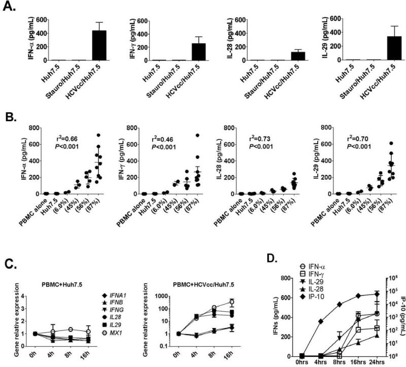
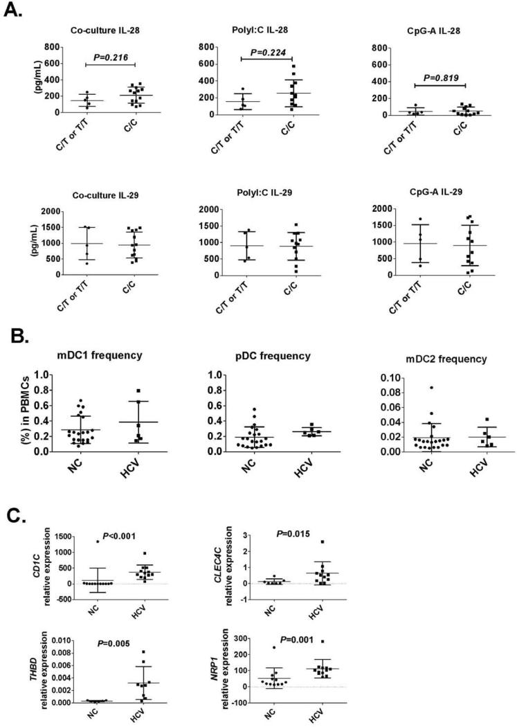
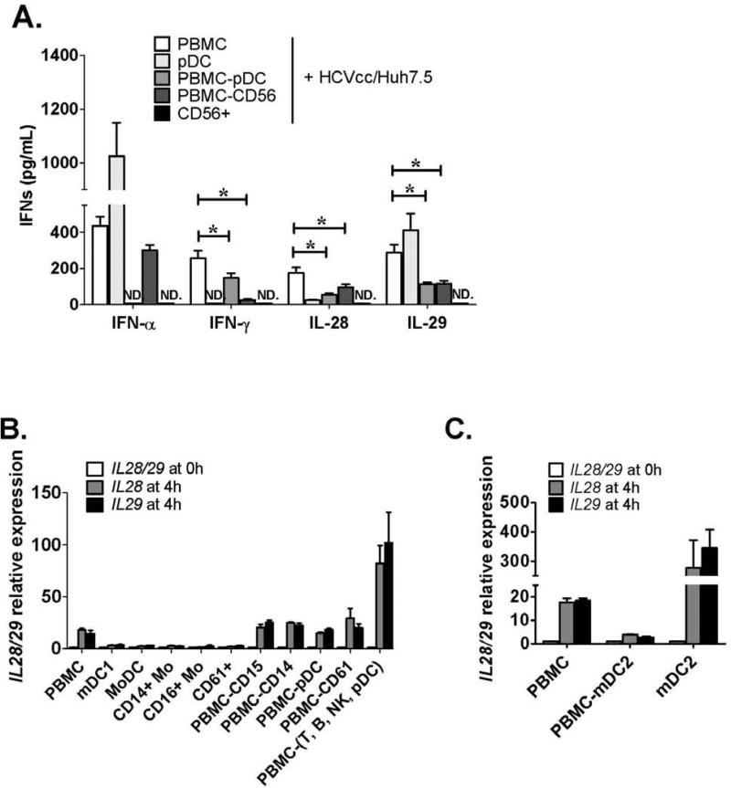
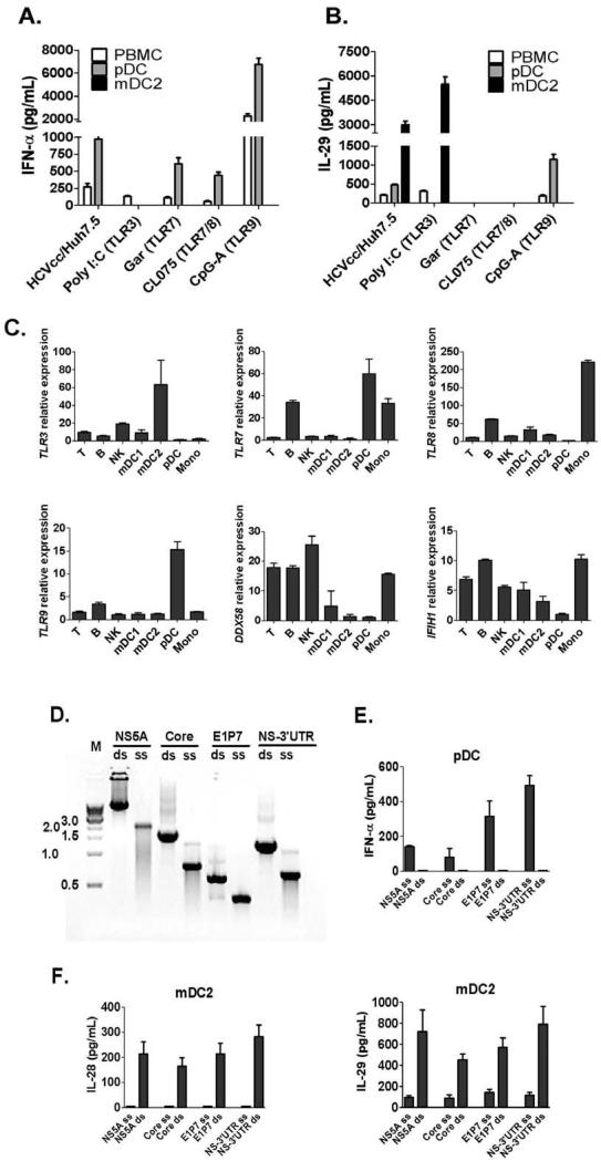
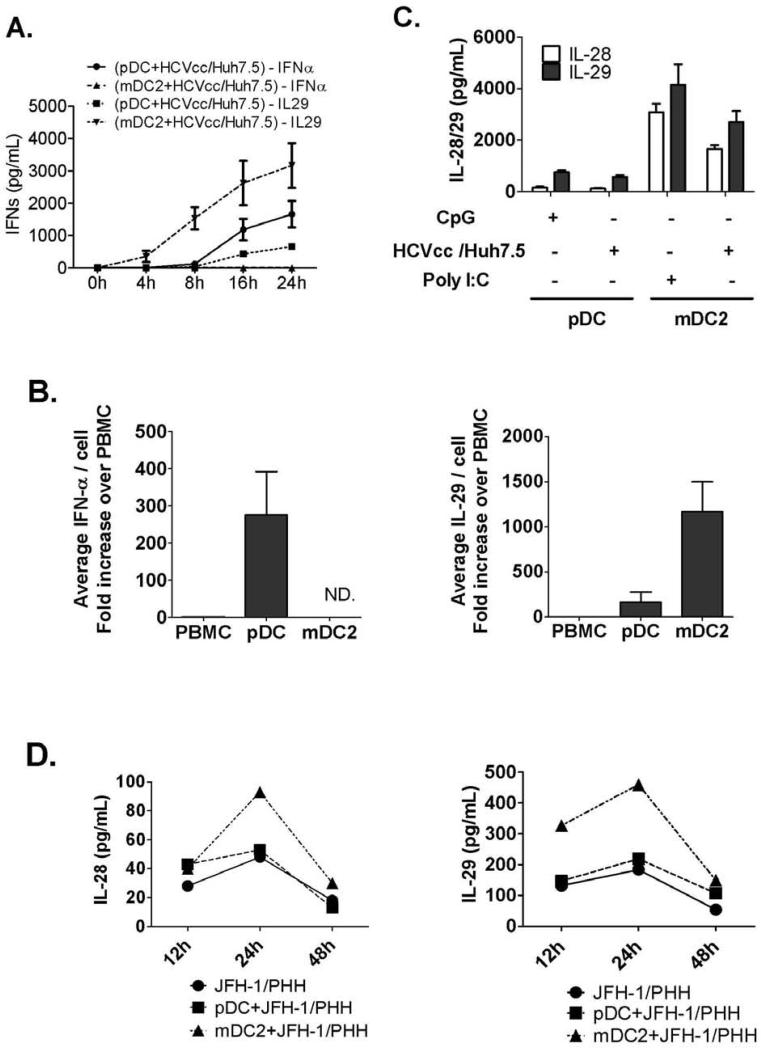
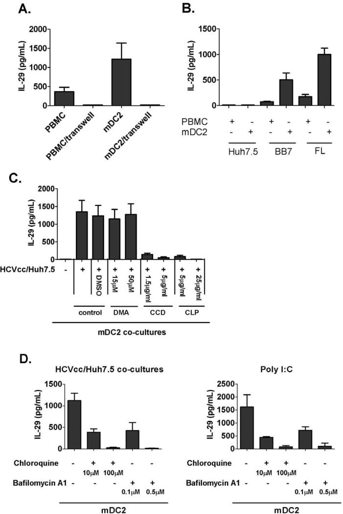
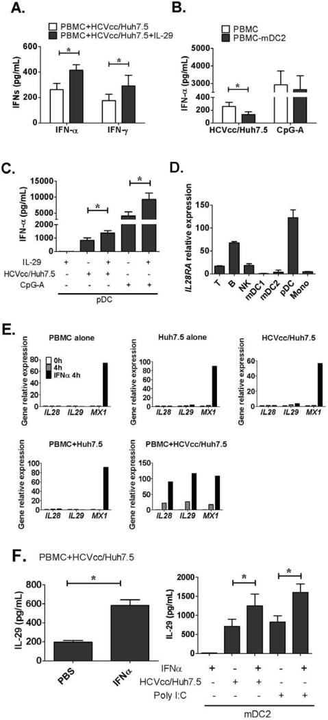
Human PBMCs recognized HCVcc/Huh7.5 cells and responded by producing IFN-α, IFN-γ, and IFN-λ. A rare subset of myeloid dendritic cells (mDCs), which are blood DC antigen (BDCA)+ (also called mDC2 cells), were the major source of IL-28 and IL-29 production in response to HCVcc/Huh7.5 cells. Plasmacytoid DCs produced IFN-α, whereas natural killer and natural killer T cells were the main source of IFN-γ production in co-culture experiments. Of the endosomal Toll-like receptors (TLRs)3, 7, 8, and 9, only TLR3 or double-stranded HCV RNA induced production of IL-28 and IL-29 by mDC2s; endosomal maturation was required. Production of IFN-α and IFN-λ were linked-IFN-λ increased production of IFN-α by plasmacytoid DCs and IFN-α significantly increased production of IFN-λ.
mDC2s are a major source of IFN-λ production by PBMCs in response to HCVcc/Huh7.5 cells. mDC2s are activated through the TLR3 pathway, indicating that human DCs efficiently can initiate an immune response against HCV infection. IFN-λ therefore has an important role in HCV infection.
III 型干扰素(IFN-λs:白细胞介素[IL]-28a、IL-28b 和 IL-29)在丙型肝炎病毒(HCV)感染中具有重要作用,但人们对哪些细胞产生这些细胞因子或如何激活其产生知之甚少。我们研究了人类免疫细胞是否能识别 HCV 感染的细胞,并通过产生 IFN-λ 做出反应。
我们用不同免疫细胞群培养健康的人外周血单核细胞(PBMC)和日本暴发性肝炎-1(JFH-1)HCV 感染的 Huh7.5(细胞培养衍生的 HCV 颗粒[HCVcc]/Huh7.5)细胞。
人 PBMC 识别 HCVcc/Huh7.5 细胞,并通过产生 IFN-α、IFN-γ 和 IFN-λ 做出反应。一种罕见的髓样树突状细胞(mDC)亚群,即血液树突状细胞抗原(BDCA)+(也称为 mDC2 细胞),是对 HCVcc/Huh7.5 细胞反应中产生 IL-28 和 IL-29 的主要来源。浆细胞样树突状细胞产生 IFN-α,而自然杀伤细胞和自然杀伤 T 细胞是共培养实验中 IFN-γ 产生的主要来源。在内体 Toll 样受体(TLR)3、7、8 和 9 中,只有 TLR3 或双链 HCV RNA 诱导 mDC2 产生 IL-28 和 IL-29;内体成熟是必需的。IFN-α 和 IFN-λ 的产生是相关联的——IFN-λ 增加浆细胞样树突状细胞产生 IFN-α,而 IFN-α 显著增加 IFN-λ 的产生。
mDC2 是 PBMC 对 HCVcc/Huh7.5 细胞反应中 IFN-λ 产生的主要来源。mDC2 通过 TLR3 途径被激活,表明人类树突状细胞能够有效地启动针对 HCV 感染的免疫反应。因此,IFN-λ 在 HCV 感染中具有重要作用。