Taams M A, Gussenhoven E J, Bos E, de Jaegere P, Roelandt J R, Sutherland G R, Bom N
Thoraxcenter, Erasmus University Rotterdam, The Netherlands.
Br Heart J. 1990 Feb;63(2):109-13. doi: 10.1136/hrt.63.2.109.
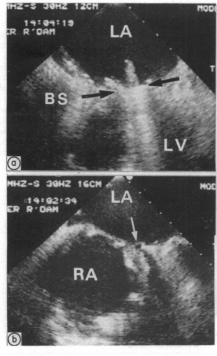
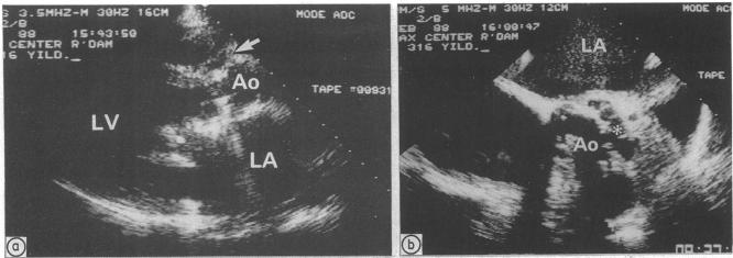
Thirty three consecutive patients with clinically suspected endocarditis were studied by both precordial cross sectional echocardiography and transoesophageal echocardiography. The diagnostic value of both techniques was assessed. The data were compared with findings at operation in 25 patients. In 21 patients with native valve endocarditis precordial echocardiography showed evidence of vegetations in six patients and suggested their presence in nine. Transoesophageal echocardiography identified vegetations in 18 patients. Complications were seen in four patients at precordial echocardiography and in nine patients at transoesophageal echocardiography. Precordial echocardiography did not show vegetations in any of the 12 patients with prosthetic valve endocarditis whereas transoesophageal echocardiography showed vegetations in four. Complications were seen in four patients at precordial echocardiography and in 10 at transoesophageal echocardiography. Echocardiographic findings were confirmed at operation in all 25 operated patients. In two patients both echocardiographic techniques had missed the perforation of the cusps of the aortic valve that was seen at operation, but this had no effect on patient management. Transoesophageal echocardiography is the best diagnostic approach when infective endocarditis is suspected in patients with either native or prosthetic valves.
对33例临床怀疑患有心内膜炎的连续患者进行了胸前区横断面超声心动图和经食管超声心动图检查。评估了这两种技术的诊断价值。将数据与25例患者的手术结果进行了比较。在21例天然瓣膜心内膜炎患者中,胸前区超声心动图显示6例有赘生物证据,9例提示有赘生物存在。经食管超声心动图在18例患者中发现了赘生物。胸前区超声心动图检查发现4例有并发症,经食管超声心动图检查发现9例有并发症。12例人工瓣膜心内膜炎患者中,胸前区超声心动图均未显示赘生物,而经食管超声心动图在4例中显示有赘生物。胸前区超声心动图检查发现4例有并发症,经食管超声心动图检查发现10例有并发症。所有25例接受手术的患者,其超声心动图检查结果在手术中均得到证实。有2例患者,两种超声心动图检查技术均未发现手术中所见的主动脉瓣瓣叶穿孔,但这对患者的治疗并无影响。当怀疑天然瓣膜或人工瓣膜患者患有感染性心内膜炎时,经食管超声心动图是最佳的诊断方法。