MR Centre of Excellence, Medical University of Vienna, Vienna, Austria.
PLoS One. 2012;7(11):e50050. doi: 10.1371/journal.pone.0050050. Epub 2012 Nov 29.
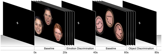
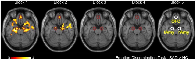
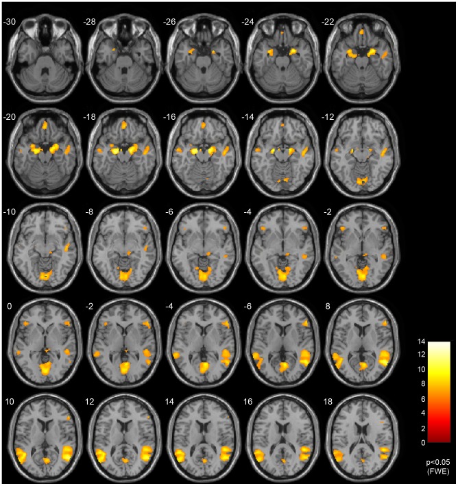
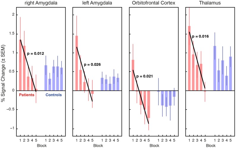
A characterizing symptom of social anxiety disorder (SAD) is increased emotional reactivity towards potential social threat in combination with impaired emotion and stress regulation. While several neuroimaging studies have linked SAD with hyperreactivity in limbic brain regions when exposed to emotional faces, little is known about habituation in both the amygdala and neocortical regulation areas. 15 untreated SAD patients and 15 age- and gender-matched healthy controls underwent functional magnetic resonance imaging during repeated blocks of facial emotion ([Formula: see text]) and object discrimination tasks ([Formula: see text]). Emotion processing networks were defined by a task-related contrast ([Formula: see text]). Linear regression was employed for assessing habituation effects in these regions. In both groups, the employed paradigm robustly activated the emotion processing and regulation network, including the amygdalae and orbitofrontal cortex (OFC). Statistically significant habituation effects were found in the amygdalae, OFC, and pulvinar thalamus of SAD patients. No such habituation was found in healthy controls. Concurrent habituation in the medial OFC and the amygdalae of SAD patients as shown in this study suggests intact functional integrity and successful short-term down-regulation of neural activation in brain areas responsible for emotion processing. Initial hyperactivation may be explained by an insufficient habituation to new stimuli during the first seconds of exposure. In addition, our results highlight the relevance of the orbitofrontal cortex in social anxiety disorders.
社交焦虑障碍(SAD)的一个特征性症状是,当暴露于情绪面孔时,对潜在社交威胁的情绪反应增强,同时情绪和应激调节受损。虽然有几项神经影像学研究将 SAD 与杏仁核和新皮层调节区域的反应过度联系起来,但对于两者的习惯化知之甚少。15 名未经治疗的 SAD 患者和 15 名年龄和性别匹配的健康对照组在重复的面部情绪([公式:见文本])和物体辨别任务([公式:见文本])期间接受功能磁共振成像。通过任务相关对比([公式:见文本])定义情绪处理网络。采用线性回归评估这些区域的习惯化效应。在两组中,所采用的范式都能强烈激活情绪处理和调节网络,包括杏仁核和眶额皮层(OFC)。在 SAD 患者的杏仁核、OFC 和丘脑枕核中发现了统计学上显著的习惯化效应。在健康对照组中没有发现这种习惯化。SAD 患者中内侧 OFC 和杏仁核的同时习惯化表明,大脑中负责情绪处理的区域具有完整的功能完整性和成功的短期神经激活下调。最初的过度激活可以通过在暴露的最初几秒钟对新刺激的适应不足来解释。此外,我们的研究结果强调了眶额皮层在社交焦虑障碍中的重要性。