• 文献检索
  • 文档翻译
  • 深度研究
  • 学术资讯
  • Suppr Zotero 插件Zotero 插件
  • 邀请有礼
  • 套餐&价格
  • 历史记录
应用&插件
Suppr Zotero 插件Zotero 插件浏览器插件Mac 客户端Windows 客户端微信小程序
定价
高级版会员购买积分包购买API积分包
服务
文献检索文档翻译深度研究API 文档MCP 服务
关于我们
关于 Suppr公司介绍联系我们用户协议隐私条款
关注我们

Suppr 超能文献

核心技术专利:CN118964589B侵权必究
粤ICP备2023148730 号-1Suppr @ 2026

文献检索

告别复杂PubMed语法,用中文像聊天一样搜索,搜遍4000万医学文献。AI智能推荐,让科研检索更轻松。

立即免费搜索

文件翻译

保留排版,准确专业,支持PDF/Word/PPT等文件格式,支持 12+语言互译。

免费翻译文档

深度研究

AI帮你快速写综述,25分钟生成高质量综述,智能提取关键信息,辅助科研写作。

立即免费体验

卵巢冠滤泡状上皮化生在一瘤牛中。

Squamous metaplasia of the rete ovarii in a Zebu cow.

机构信息

Departamento de Clínica e Cirurgia Veterinárias, Escola de Veterinária, Universidade Federal de Minas Gerais, Minas Gerais, Brasil.

出版信息

BMC Vet Res. 2012 Dec 5;8:235. doi: 10.1186/1746-6148-8-235.

DOI:10.1186/1746-6148-8-235
PMID:23217175
原文链接:https://pmc.ncbi.nlm.nih.gov/articles/PMC3528438/
Abstract

BACKGROUND

Stratified keratinizing squamous epithelium in the ovary has been associated with the diagnosis of ovarian teratoma in cows. Recently, the diagnosis of "epidermoid cyst" has been proposed. A case of squamous metaplasia of the rete ovarii in a Zebu cow is described in this report.

CASE PRESENTATION

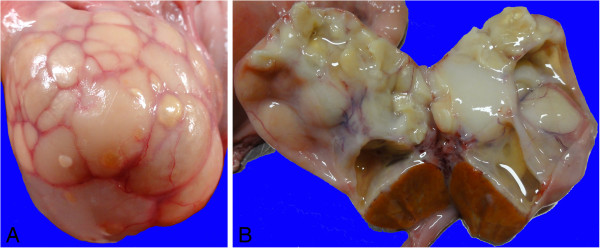
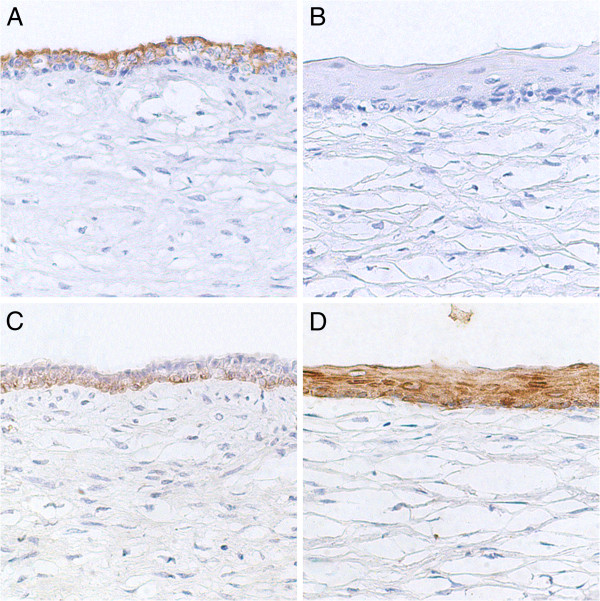
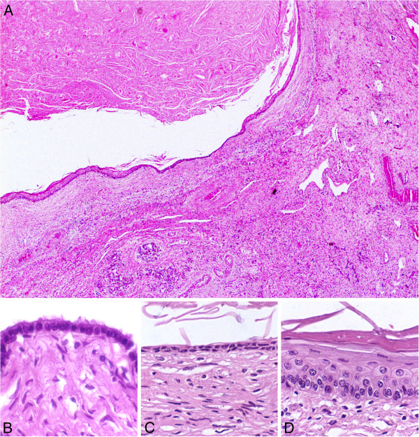
A crossbreed Zebu cow had both ovaries enlarged with multiple cysts. Most cysts were lined by well differentiated keratinizing stratified squamous epithelium and filled with keratinized lamellar material. Some cysts were lined by an epithelial layer that ranged from single cuboidal, double cuboidal epithelium, stratified non keratinized epithelium, and areas of keratinizing stratified squamous epithelium. Single or double layered cuboidal epithelia of the cysts expressed low molecular weight cytokeratin 7, whose expression was absent in the keratinizing stratified squamous epithelia of same cysts. Conversely, high molecular weight cytokeratins 1, 5, 10, and 14 were strongly expressed by the keratinizing stratified epithelium.

CONCLUSION

Squamous metaplasia of the rete ovarii was diagnosed. Squamous metaplasia of the rete ovarii, may account for some of the previously described squamous lesions in the ovary, which may have been misinterpreted as teratoma or epidermoid cysts.

摘要

背景

卵巢中分层角化的鳞状上皮与牛卵巢畸胎瘤的诊断有关。最近,提出了“表皮样囊肿”的诊断。本文报道了一例努比亚奶牛卵巢网腔的鳞状化生。

病例介绍

杂交努比亚奶牛的两个卵巢均增大,伴有多个囊肿。大多数囊肿由分化良好的角化分层鳞状上皮衬里,并充满角化的板层物质。一些囊肿衬有上皮层,范围从单层立方上皮、双层立方上皮、非角化分层上皮和角化分层鳞状上皮区域。囊肿的单层或双层立方上皮表达低分子量细胞角蛋白 7,而同一囊肿的角化分层鳞状上皮中则没有表达。相反,高分子量细胞角蛋白 1、5、10 和 14 在角化分层上皮中强烈表达。

结论

诊断为卵巢网腔的鳞状化生。卵巢网腔的鳞状化生可能是以前描述的卵巢中一些鳞状病变的原因,这些病变可能被误诊为畸胎瘤或表皮样囊肿。

https://cdn.ncbi.nlm.nih.gov/pmc/blobs/856b/3528438/7b688d64c223/1746-6148-8-235-3.jpg
https://cdn.ncbi.nlm.nih.gov/pmc/blobs/856b/3528438/01db4b8a760d/1746-6148-8-235-1.jpg
https://cdn.ncbi.nlm.nih.gov/pmc/blobs/856b/3528438/0b606688a07c/1746-6148-8-235-2.jpg
https://cdn.ncbi.nlm.nih.gov/pmc/blobs/856b/3528438/7b688d64c223/1746-6148-8-235-3.jpg
https://cdn.ncbi.nlm.nih.gov/pmc/blobs/856b/3528438/01db4b8a760d/1746-6148-8-235-1.jpg
https://cdn.ncbi.nlm.nih.gov/pmc/blobs/856b/3528438/0b606688a07c/1746-6148-8-235-2.jpg
https://cdn.ncbi.nlm.nih.gov/pmc/blobs/856b/3528438/7b688d64c223/1746-6148-8-235-3.jpg

相似文献

1
Squamous metaplasia of the rete ovarii in a Zebu cow.卵巢冠滤泡状上皮化生在一瘤牛中。
BMC Vet Res. 2012 Dec 5;8:235. doi: 10.1186/1746-6148-8-235.
2
Bilateral ovarian cysts originating from rete ovarii in an African green monkey (Cercopithecus aethiops).一只非洲绿猴(猕猴属埃塞俄比亚种)双侧卵巢囊肿起源于卵巢网。
J Vet Med Sci. 2012 Sep;74(9):1229-32. doi: 10.1292/jvms.11-0354. Epub 2012 May 21.
3
Feline cystic rete ovarii.猫卵巢囊状网
Vet Pathol. 1984 May;21(3):304-7. doi: 10.1177/030098588402100307.
4
Ovarian cysts in MRL / MpJ mice are derived from the extraovarian rete: a developmental study.MRL/MpJ 小鼠的卵巢囊肿来源于卵巢外的 rete 组织:一项发育研究。
J Anat. 2011 Dec;219(6):743-55. doi: 10.1111/j.1469-7580.2011.01431.x. Epub 2011 Sep 26.
5
Three cases of ovarian epidermoid cysts in cattle.
Vet Pathol. 2002 Nov;39(6):744-6. doi: 10.1354/vp.39-6-744.
6
Cystic rete ovarii and uterine tube adenoma in a rabbit.兔的卵巢囊肿性网织细胞瘤和输卵管腺瘤
J Vet Med Sci. 2014 Jun;76(6):909-12. doi: 10.1292/jvms.14-0053. Epub 2014 Feb 27.
7
Ovarian cysts in MRL/MpJ mice originate from rete ovarii.MRL/MpJ小鼠的卵巢囊肿起源于卵巢网。
Anat Histol Embryol. 2007 Jun;36(3):172-8. doi: 10.1111/j.1439-0264.2006.00728.x.
8
Mast cells and Kurloff cells - Their detection throughout the oestrous cycle in normal guinea pig ovaries and in guinea pigs with cystic rete ovarii.肥大细胞和库尔洛夫细胞——在正常豚鼠卵巢和患有囊状卵巢网的豚鼠整个发情周期中的检测。
Res Vet Sci. 2021 May;136:512-518. doi: 10.1016/j.rvsc.2021.04.003. Epub 2021 Apr 14.
9
Quantitative trait locus analysis of ovarian cysts derived from rete ovarii in MRL/MpJ mice.MRL/MpJ 小鼠卵巢冠来源的卵巢囊肿的数量性状位点分析。
Mamm Genome. 2010 Apr;21(3-4):162-71. doi: 10.1007/s00335-010-9254-x. Epub 2010 Feb 25.
10
E-cadherin expression and bromodeoxyuridine incorporation during development of ovarian inclusion cysts in age-matched breeder and incessantly ovulated CD-1 mice.年龄匹配的繁殖期和持续排卵的CD-1小鼠卵巢包涵囊肿发育过程中E-钙黏蛋白的表达及溴脱氧尿苷掺入情况。
Reprod Biol Endocrinol. 2007 Apr 11;5:14. doi: 10.1186/1477-7827-5-14.

引用本文的文献

1
Rediscovering the , a secreting auxiliary structure to the ovary.重新发现卵巢的一个分泌性附属结构。 不过你提供的原文“Rediscovering the, a secreting auxiliary structure to the ovary.”似乎不太完整,有个逗号位置不太对,可能会影响更准确理解。
Elife. 2025 Mar 19;13:RP96662. doi: 10.7554/eLife.96662.
2
Rediscovering the : a secreting auxiliary structure to the ovary.重新发现:一种卵巢的分泌性辅助结构。 (注:原句中“Rediscovering the :”表述不完整,这里只能按现有内容尽量准确翻译)
bioRxiv. 2024 Nov 11:2023.11.08.566085. doi: 10.1101/2023.11.08.566085.

本文引用的文献

1
Squamous lesions of the ovary.卵巢的鳞状病变。
Arch Pathol Lab Med. 2011 Dec;135(12):1611-4. doi: 10.5858/arpa.2010-0640-RS.
2
Ovarian and paraovarian squamous-lined cysts (epidermoid cysts): a clinicopathologic study of 18 cases with comparison to mature cystic teratomas.卵巢及卵巢旁衬覆鳞状上皮的囊肿(表皮样囊肿):18例临床病理研究并与成熟囊性畸胎瘤对比
Int J Gynecol Pathol. 2009 Mar;28(2):193-6. doi: 10.1097/PGP.0b013e318187e147.
3
Three cases of ovarian epidermoid cysts in cattle.
Vet Pathol. 2002 Nov;39(6):744-6. doi: 10.1354/vp.39-6-744.
4
Structural and cell surface antigen expression in the rete ovarii and epoophoron differs from that in the Fallopian tube and in endometriosis.卵巢网和卵巢冠中的结构及细胞表面抗原表达与输卵管及子宫内膜异位症中的不同。
Histopathology. 2000 Jul;37(1):64-9. doi: 10.1046/j.1365-2559.2000.00938.x.
5
Cystic corpora lutea and normal and cystic graafian follicles in the cow.
Aust Vet J. 1968 Jul;44(7):304-8. doi: 10.1111/j.1751-0813.1968.tb09104.x.