Chang Jeannette, Arbeláez Pablo, Switz Neil, Reber Clay, Tapley Asa, Davis J Lucian, Cattamanchi Adithya, Fletcher Daniel, Malik Jitendra
UC Berkeley, Department of Electrical Engineering and Computer Sciences, USA.
Med Image Comput Comput Assist Interv. 2012;15(Pt 3):345-52. doi: 10.1007/978-3-642-33454-2_43.
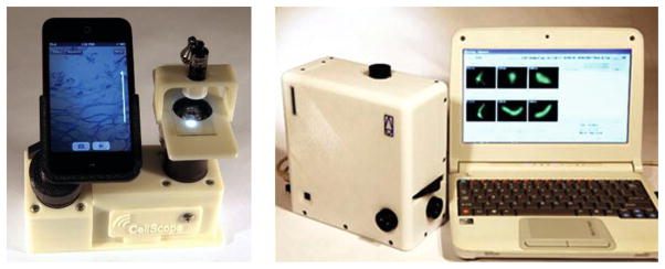
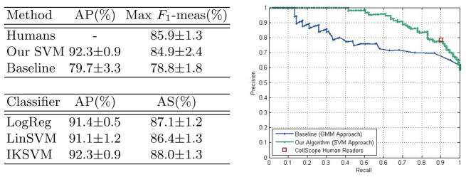
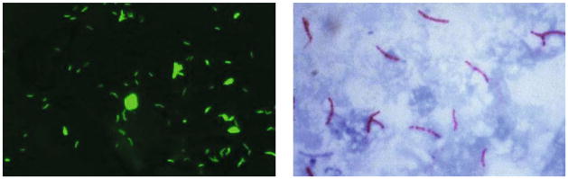
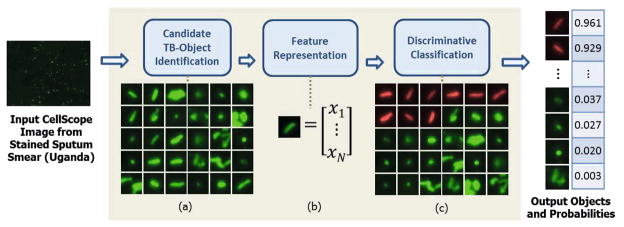
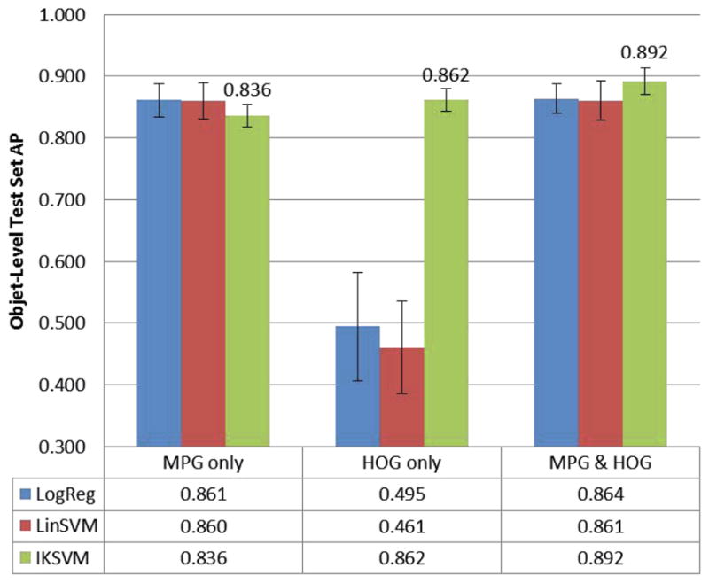
In low-resource areas, the most common method of tuberculosis (TB) diagnosis is visual identification of rod-shaped TB bacilli in microscopic images of sputum smears. We present an algorithm for automated TB detection using images from digital microscopes such as CellScope, a novel, portable device capable of brightfield and fluorescence microscopy. Automated processing on such platforms could save lives by bringing healthcare to rural areas with limited access to laboratory-based diagnostics. Our algorithm applies morphological operations and template matching with a Gaussian kernel to identify candidate TB-objects. We characterize these objects using Hu moments, geometric and photometric features, and histograms of oriented gradients and then perform support vector machine classification. We test our algorithm on a large set of CellScope images (594 images corresponding to 290 patients) from sputum smears collected at clinics in Uganda. Our object-level classification performance is highly accurate, with average precision of 89.2% +/- 2.1%. For slide-level classification, our algorithm performs at the level of human readers, demonstrating the potential for making a significant impact on global healthcare.
在资源匮乏地区,结核病(TB)诊断的最常见方法是在痰涂片显微镜图像中目视识别杆状结核杆菌。我们提出了一种使用来自数字显微镜(如CellScope,一种新型便携式设备,能够进行明场和荧光显微镜检查)的图像进行结核病自动检测的算法。在此类平台上进行自动化处理可以通过将医疗服务带到难以获得基于实验室诊断的农村地区来挽救生命。我们的算法应用形态学操作和高斯核模板匹配来识别候选结核物体。我们使用Hu矩、几何和光度特征以及定向梯度直方图对这些物体进行特征描述,然后进行支持向量机分类。我们在乌干达诊所收集的大量CellScope痰涂片图像(594幅图像,对应290名患者)上测试了我们的算法。我们的物体级分类性能高度准确,平均精度为89.2%±2.1%。对于玻片级分类,我们的算法表现与人类读者相当,证明了其对全球医疗保健产生重大影响的潜力。