Center on Aging Psychology, Key Laboratory of Mental Health, Institute of Psychology, Chinese Academy of Sciences, Beijing, China.
PLoS One. 2013;8(1):e53148. doi: 10.1371/journal.pone.0053148. Epub 2013 Jan 2.
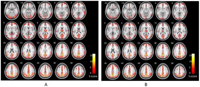
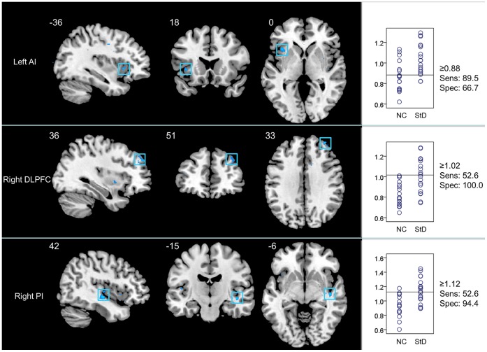
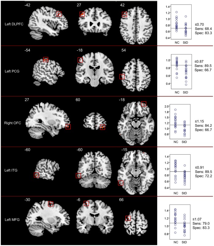
The early detection of major depression in elderly individuals who are at risk of developing the disease is of prime importance when it comes to the prevention of geriatric depression. We used resting-state functional magnetic resonance imaging (fMRI) to examine changes in regional homogeneity (ReHo) of spontaneous activity in late-life subthreshold depression (StD), and we evaluated the sensitivity/specificity performance of these changes. Nineteen elderly individuals with StD and 18 elderly controls underwent a resting-state fMRI scan. The ReHo approach was employed to examine whether StD was related to alterations in resting-state neural activity, in the form of abnormal regional synchronization. Receiver operating characteristic curve analysis and the Fisher stepwise discriminant analysis were used to evaluate the sensitivity/specificity characteristics of the ReHo index in discriminating between the StD subjects and normal controls. The results demonstrated that, compared to controls, StD subjects display lower ReHo in the right orbitofrontal cortex (OFC), left dorsolateral prefrontal cortex (DLPFC), left postcentral gyrus (PCG), and left middle frontal and inferior temporal gyri, as well as higher ReHo in the bilateral insula and right DLPFC. The left PCG and the right DLPFC, OFC, and posterior insula, together reported a predictive accuracy of 91.9%. These results suggest that the regional activity coherence was changed in the resting brain of StD subjects, and that these alterations may serve as potential markers for the early detection of StD in late-life depression.
在预防老年抑郁症方面,早期发现有患病风险的老年个体中是否存在重度抑郁症至关重要。我们使用静息态功能磁共振成像(fMRI)来检查晚年亚临床抑郁(StD)患者自发活动的局部一致性(ReHo)变化,并评估这些变化的敏感性/特异性表现。19 名患有 StD 的老年患者和 18 名老年对照组接受了静息态 fMRI 扫描。采用 ReHo 方法来研究 StD 是否与静息状态下的神经活动改变有关,这种改变表现为异常的区域同步化。采用接收者操作特征曲线分析和 Fisher 逐步判别分析来评估 ReHo 指数在区分 StD 患者和正常对照组方面的敏感性/特异性特征。结果表明,与对照组相比,StD 患者右侧眶额皮质(OFC)、左侧背外侧前额叶皮质(DLPFC)、左侧中央后回(PCG)和左侧额中回和颞下回的 ReHo 降低,双侧岛叶和右侧 DLPFC 的 ReHo 升高。左侧 PCG 和右侧 DLPFC、OFC 和后岛叶联合报告的预测准确率为 91.9%。这些结果表明,StD 患者静息脑的局部活动连贯性发生了改变,这些改变可能是早期发现老年抑郁症中 StD 的潜在标志物。