Immune Imaging Program, The Centenary Institute Newtown, NSW, Australia ; Sydney Medical School, University of Sydney Sydney, NSW, Australia.
Front Cell Neurosci. 2013 Jan 8;6:67. doi: 10.3389/fncel.2012.00067. eCollection 2012.
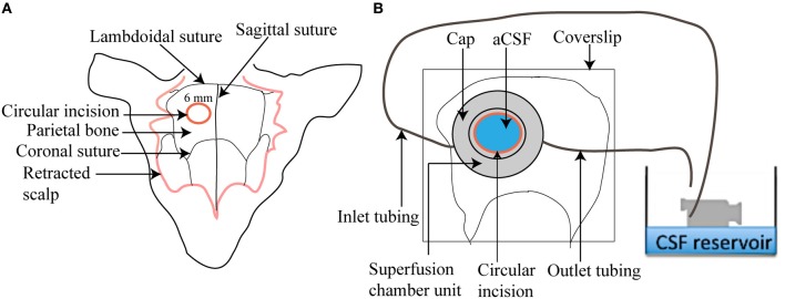
Intravital imaging of the superficial brain tissue in mice represents a powerful tool for the dissection of the cellular and molecular cues underlying inflammatory and infectious central nervous system (CNS) diseases. We present here a step-by-step protocol that will enable a non-specialist to set up a two-photon brain-imaging model. The protocol offers a two-part approach that is specifically optimized for imaging leukocytes but can be easily adapted to answer varied CNS-related biological questions. The protocol enables simultaneous visualization of fluorescently labeled immune cells, the pial microvasculature and extracellular structures such as collagen fibers at high spatial and temporal resolution. Intracranial structures are exposed through a cranial window, and physiologic conditions are maintained during extended imaging sessions via continuous superfusion of the brain surface with artificial cerebrospinal fluid (aCSF). Experiments typically require 1-2 h of preparation, which is followed by variable periods of immune cell tracking. Our methodology converges the experience of two laboratories over the past 10 years in diseased animal models such as cerebral ischemia, lupus, cerebral malaria, and toxoplasmosis. We exemplify the utility of this protocol by tracking leukocytes in transgenic mice in the pial vessels under steady-state conditions.
活体小鼠脑部组织成像技术是研究炎症和感染性中枢神经系统(CNS)疾病中细胞和分子信号的有力工具。本方案详细介绍了一套标准化流程,可使非专业人员建立双光子脑成像模型。该方案提供了一种两步法,专门针对白细胞成像进行了优化,但也可以轻松调整以回答各种与 CNS 相关的生物学问题。该方案可实现对荧光标记免疫细胞、软脑膜微血管和细胞外结构(如胶原纤维)的高时空分辨率同时可视化。通过颅窗暴露颅内结构,并通过持续向脑表面灌注人工脑脊液(aCSF)在延长的成像过程中维持生理条件。实验通常需要 1-2 小时的准备时间,然后根据免疫细胞的追踪时间而定。本方法结合了过去 10 年中两个实验室在脑缺血、狼疮、脑疟疾和弓形体病等疾病动物模型中的经验。我们通过在稳定状态下追踪转基因小鼠软脑膜血管中的白细胞来举例说明该方案的实用性。