Microscopy Core Facility, Max Planck Institute for Infection Biology Berlin, Germany.
Front Immunol. 2013 Jan 9;3:413. doi: 10.3389/fimmu.2012.00413. eCollection 2012.
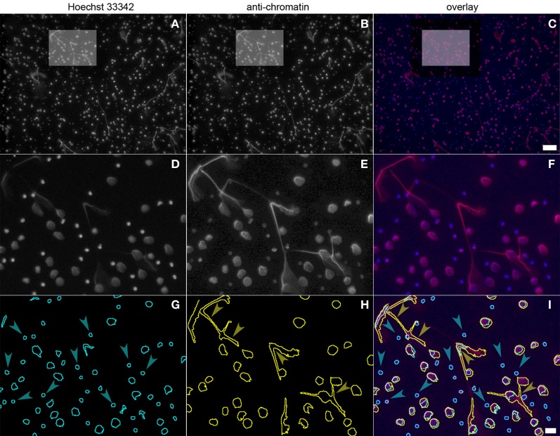
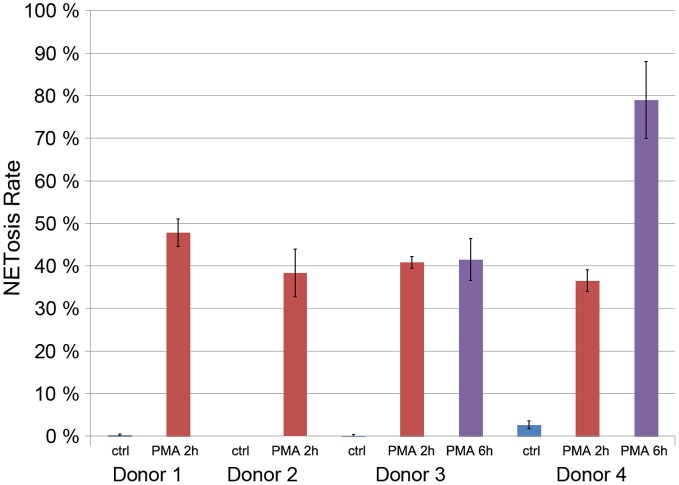
Neutrophil Extracellular Traps (NETs) consist of decondensed chromatin studded with granular and some cytoplasmic proteins. They are formed by activated neutrophil granulocytes, also called polymorphonuclear leukocytes (PMN) as the result of an active cell death program, named NETosis. NETosis can be induced by a wide range of stimuli including coculture of neutrophils with pathogens (bacteria, fungi, parasites, virus particles), activated platelets, or pathogen components. The first step of the NETotic cascade is stimulation of one or several receptors followed by activation of the Raf/MEK/ERK pathway that culminates in the assembly of the multimeric NADPH oxidase complex and the production of reactive oxygen species (ROS). Later, intracellular membranes disintegrate, the granular protein Neutrophil Elastase enters the nucleus and processes core histones that also get hypercitrullinated. This leads to decondensation and mobilization of chromatin. The amount of NET formation varies with the degree of stimulation, and this is dependent on the type and concentration of the stimulus. NETs can be quantified using various methods including fluorescence microscopy or measuring DNA release. Each of these methods have specific drawbacks: analysis of fluorescence microscopy is prone to subjective variations, and DNA release does not differentiate between DNA that has been released by NETosis or by other forms of cell death. Here we present a protocol to semi-automatically quantify NET formation. It relies on the observation that anti-chromatin antibodies bind more readily to decondensed chromatin present in the nuclei of cells undergoing NETosis and in the NETs. Relating the fluorescence signals of the anti-chromatin antibody to the signals of a DNA-binding dye allows the automatic calculation of the percentage of netting neutrophils. This method does not require sophisticated microscopic equipment, and the images are quantified with a public-domain software package.
中性粒细胞胞外诱捕网(NETs)由带有颗粒和一些细胞质蛋白的去凝聚染色质组成。它们是由活化的中性粒细胞粒细胞形成的,也称为多形核白细胞(PMN),是一种称为 NETosis 的活性细胞死亡程序的结果。NETosis 可由多种刺激物诱导,包括与病原体(细菌、真菌、寄生虫、病毒颗粒)、活化的血小板或病原体成分共培养的中性粒细胞。NETotic 级联的第一步是刺激一个或多个受体,随后激活 Raf/MEK/ERK 途径,最终导致多聚体 NADPH 氧化酶复合物的组装和活性氧物质(ROS)的产生。随后,细胞内膜解体,颗粒蛋白弹性蛋白酶进入细胞核并处理核心组蛋白,这些组蛋白也被过度瓜氨酸化。这导致染色质去凝聚和动员。NET 形成的数量随刺激程度而变化,这取决于刺激的类型和浓度。NET 可以使用各种方法进行定量,包括荧光显微镜或测量 DNA 释放。这些方法各有特定的缺点:荧光显微镜分析容易受到主观变化的影响,而 DNA 释放不能区分 NETosis 或其他形式的细胞死亡释放的 DNA。在这里,我们提出了一种半自动定量 NET 形成的方法。它依赖于这样一种观察,即抗染色质抗体更容易与正在经历 NETosis 的细胞的细胞核中和 NETs 中存在的去凝聚染色质结合。将抗染色质抗体的荧光信号与 DNA 结合染料的信号相关联,允许自动计算出现 NET 的中性粒细胞的百分比。该方法不需要复杂的显微镜设备,并且使用公共域软件包对图像进行定量。