DTU Nanotech, Technical University of Denmark, Lyngby, Denmark.
PLoS One. 2013;8(1):e53307. doi: 10.1371/journal.pone.0053307. Epub 2013 Jan 9.
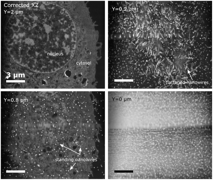
Using high resolution focused ion beam scanning electron microscopy (FIB-SEM) we study the details of cell-nanostructure interactions using serial block face imaging. 3T3 Fibroblast cellular monolayers are cultured on flat glass as a control surface and on two types of nanostructured scaffold substrates made from silicon black (Nanograss) with low- and high nanowire density. After culturing for 72 hours the cells were fixed, heavy metal stained, embedded in resin, and processed with FIB-SEM block face imaging without removing the substrate. The sample preparation procedure, image acquisition and image post-processing were specifically optimised for cellular monolayers cultured on nanostructured substrates. Cells display a wide range of interactions with the nanostructures depending on the surface morphology, but also greatly varying from one cell to another on the same substrate, illustrating a wide phenotypic variability. Depending on the substrate and cell, we observe that cells could for instance: break the nanowires and engulf them, flatten the nanowires or simply reside on top of them. Given the complexity of interactions, we have categorised our observations and created an overview map. The results demonstrate that detailed nanoscale resolution images are required to begin understanding the wide variety of individual cells' interactions with a structured substrate. The map will provide a framework for light microscopy studies of such interactions indicating what modes of interactions must be considered.
我们使用高分辨率聚焦离子束扫描电子显微镜(FIB-SEM)通过连续块面成像研究细胞-纳米结构相互作用的细节。3T3 成纤维细胞单层培养在平板玻璃上作为对照表面和两种由硅黑(Nanograss)制成的纳米结构支架基底上,这两种纳米结构支架基底的纳米线密度分别较低和较高。培养 72 小时后,将细胞固定、重金属染色、嵌入树脂,并在不去除基底的情况下进行 FIB-SEM 块面成像处理。特别优化了用于在纳米结构化基底上培养的细胞单层的样品制备程序、图像采集和图像后处理。细胞与纳米结构之间的相互作用范围广泛,这取决于表面形态,但在同一基底上的不同细胞之间也存在很大差异,说明了广泛的表型可变性。根据基底和细胞的不同,我们观察到细胞可以:例如,折断纳米线并吞噬它们,将纳米线压平或只是位于它们之上。鉴于相互作用的复杂性,我们对我们的观察结果进行了分类,并创建了一个概述图。结果表明,需要详细的纳米级分辨率图像来开始理解各种细胞与结构化基底的相互作用。该地图将为这种相互作用的明场显微镜研究提供一个框架,指出必须考虑哪些相互作用模式。