School of Food Science and Pharmacy, Zhejiang Provincial Key Engineering Technology Research Center of Marine Biomedical Products, Zhejiang Ocean University, Zhoushan 316000, China.
Mar Drugs. 2013 Jan 23;11(1):266-73. doi: 10.3390/md11010266.
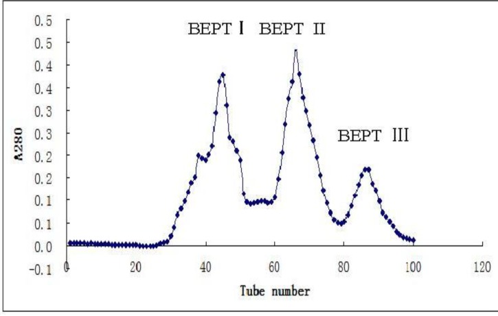
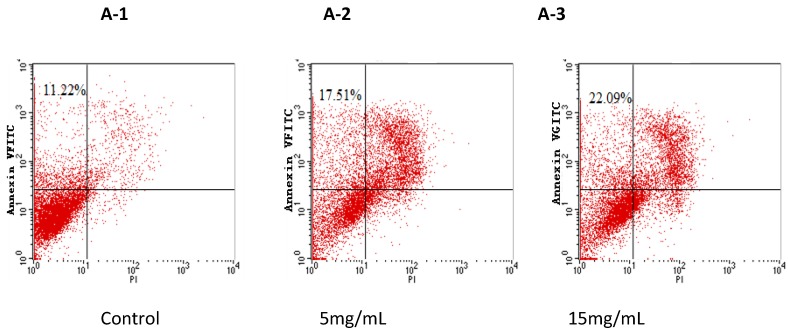
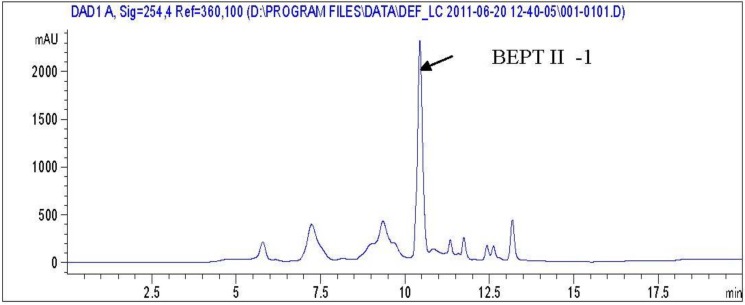
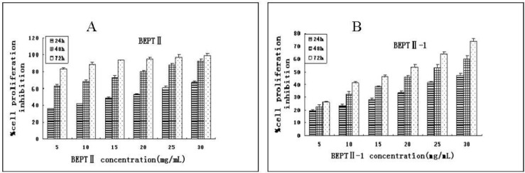
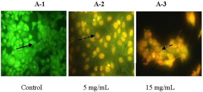
Bullacta exarata was hydrolyzed with trypsin to prepare peptides; Hydrolysates were isolated by ultrafiltration and purified using G-25 gel filtration. The purity of the Bullacta exarata was demonstrated by HPLC and its peptide sequence analysis was detected. The effects of BEPT II and BEPT II-1 on the proliferation of PC-3 cells were examined using a MTT assay. BEPT II and BEPT II-1 significantly inhibited the proliferation of PC-3 cells in a time- and dose-dependent manner. Annexin V/PI double staining studies showed exposing PC-3 cells to 5, or 15 mg/mL BEPT II-1 for 24 h increased the percentage of the early stage of apoptotic cells from 11.22% to 22.09%. In addition, typical morphologic changes were observed in the cells with acridine orange/ethidium bromide staining. These data support that BEPT II-1 has anticancer properties and merits further investigation to understand the mechanisms of BEPT II-1-induced apoptosis in PC-3 cells.
海兔经胰蛋白酶水解制备得到肽;通过超滤法对水解产物进行分离,再利用 G-25 凝胶过滤进行纯化。通过 HPLC 验证海兔肽的纯度,并检测其肽序列分析。采用 MTT 法检测 BEPT II 和 BEPT II-1 对 PC-3 细胞增殖的影响。BEPT II 和 BEPT II-1 呈时间和剂量依赖性显著抑制 PC-3 细胞的增殖。Annexin V/PI 双重染色研究表明,将 PC-3 细胞暴露于 5 或 15 mg/mL BEPT II-1 24 h 后,早期凋亡细胞的比例从 11.22%增加到 22.09%。此外,吖啶橙/溴化乙锭染色观察到细胞出现典型的形态变化。这些数据表明 BEPT II-1 具有抗癌特性,值得进一步研究以了解 BEPT II-1 诱导 PC-3 细胞凋亡的机制。