Suppr超能文献

Fuchs 内皮角膜营养不良:基于中央至周边厚度比的主观分级与客观分级。

Fuchs' endothelial corneal dystrophy: subjective grading versus objective grading based on the central-to-peripheral thickness ratio.

机构信息

Department of Ophthalmology, Mayo Clinic, Rochester, Minnesota 55905, USA.

出版信息

Ophthalmology. 2013 Apr;120(4):687-94. doi: 10.1016/j.ophtha.2012.09.022. Epub 2013 Jan 28.

Abstract

PURPOSE

To assess interobserver agreement between 2 corneal specialists grading Fuchs' dystrophy clinically and to determine if the corneal central-to-peripheral thickness ratio (CPTR) may be an alternative and objective metric of disease severity.

DESIGN

Cross-sectional study.

PARTICIPANTS

Forty-five eyes (26 subjects) with mild and moderate Fuchs' dystrophy, 73 eyes (60 subjects) with advanced Fuchs' dystrophy, and 267 eyes (142 subjects) with normal corneas.

METHODS

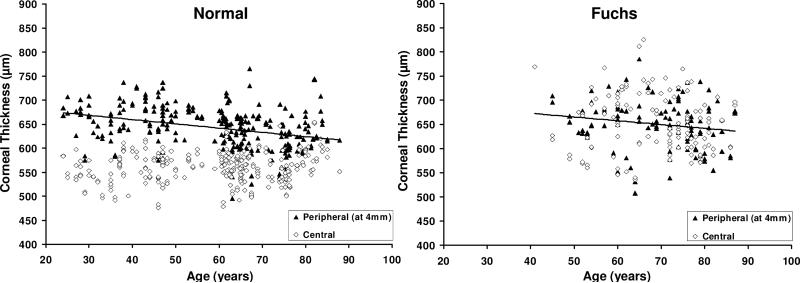
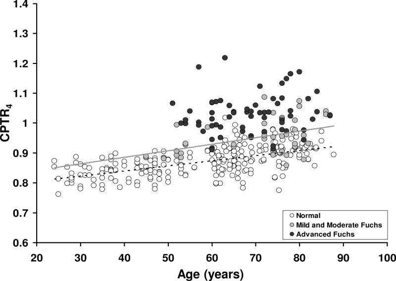
Corneas with Fuchs' dystrophy were graded by 2 corneal specialists based on the confluence and area of guttae and the presence or absence of edema. Central corneal thickness (CCT) and peripheral corneal thickness at 4 mm from the center (PCT4) were measured by using scanning-slit pachymetry. The value of CPTR4 was the quotient of CCT and PCT4.

MAIN OUTCOME MEASURES

Interobserver agreement for clinical grade and CPTR4.

RESULTS

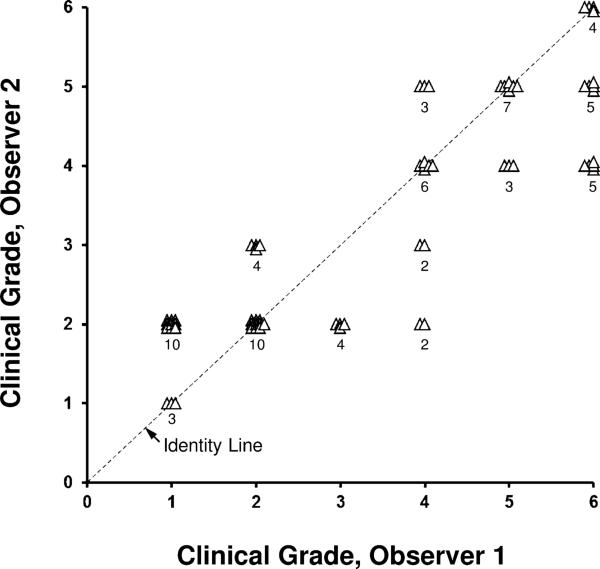
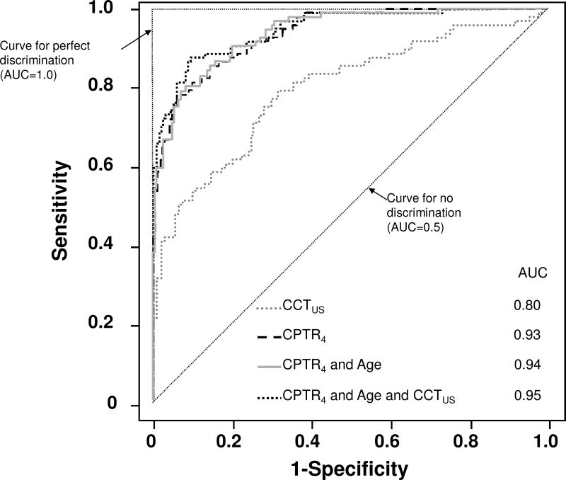
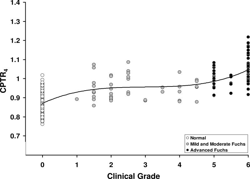
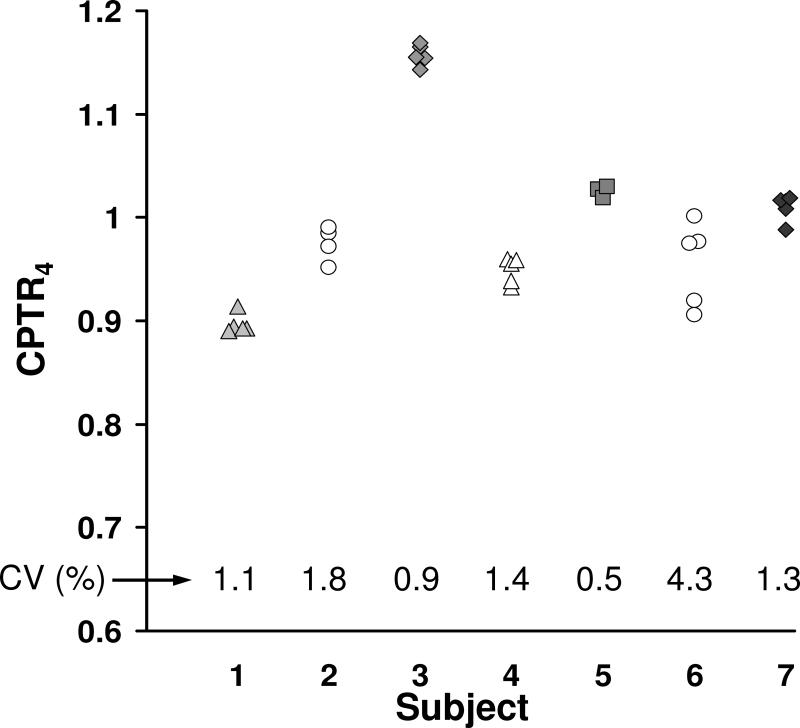
Interobserver agreement for clinical grading of Fuchs' dystrophy was moderate (κ = 0.32; 95% confidence interval, 0.19-0.45). In normal corneas, CCT was not correlated with age (r = -0.10; P = 0.28; n = 267), PCT4 decreased with age (r = -0.33; P<0.001; n = 254), and CPTR4 increased with age (r = 0.59; P<0.001; n = 254). Central corneal thickness was higher in Fuchs' dystrophy (652 ± 61 μm; n = 118) than in normal corneas (559 ± 31 μm; n = 267; P<0.001). Also, PCT4 was higher in Fuchs' dystrophy (650 ± 51 μm; n = 107) than in normal corneas (643 ± 43 μm; n = 254; P<0.001 after adjusting thickness for age). Furthermore, CPTR4 was higher in advanced Fuchs' dystrophy (1.03 ± 0.07; n = 65) than in mild and moderate Fuchs' dystrophy (0.95 ± 0.07; n = 42; age-adjusted P<0.001), which in turn was higher than in normal corneas (0.87 ± 0.05; n = 254; age-adjusted P<0.001). Finally, CPTR4 was highly correlated with clinical grade of Fuchs' dystrophy (r = 0.77; P<0.001; n = 361), was repeatable (median coefficient of variation, 1.3%), and provided excellent discrimination between Fuchs' dystrophy and normal corneas (area under the receiver operator characteristic curve, 0.93).

CONCLUSIONS

Agreement between corneal specialists for the subjective and morphologic clinical grading of Fuchs' dystrophy is only moderate. The corneal CPTR is an objective, repeatable, and possibly functional, metric of severity of Fuchs' dystrophy that warrants further investigation to determine its role in monitoring disease progression and predicting the need for keratoplasty.

摘要

目的

评估 2 位角膜专家在临床分级 Fuchs 角膜营养不良方面的观察者间一致性,并确定角膜中央-周边厚度比(CPTR)是否可以作为疾病严重程度的替代和客观指标。

设计

横断面研究。

参与者

45 只眼(26 例)患有轻度和中度 Fuchs 角膜营养不良,73 只眼(60 例)患有晚期 Fuchs 角膜营养不良,267 只眼(142 例)患有正常角膜。

方法

使用角膜地形图仪,2 位角膜专家根据角膜滴状混浊的融合和面积以及是否存在水肿来对角膜进行分级。中央角膜厚度(CCT)和距中心 4mm 处的周边角膜厚度(PCT4)通过扫描狭缝式角膜测厚仪进行测量。CPTR4 的值为 CCT 与 PCT4 的商。

主要观察指标

临床分级和 CPTR4 的观察者间一致性。

结果

Fuchs 角膜营养不良临床分级的观察者间一致性为中度(κ=0.32;95%置信区间,0.19-0.45)。在正常角膜中,CCT 与年龄无相关性(r=-0.10;P=0.28;n=267),PCT4 随年龄下降(r=-0.33;P<0.001;n=254),CPTR4 随年龄增加(r=0.59;P<0.001;n=254)。Fuchs 角膜营养不良的 CCT(652±61μm;n=118)高于正常角膜(559±31μm;n=267;P<0.001)。此外,Fuchs 角膜营养不良的 PCT4(650±51μm;n=107)高于正常角膜(643±43μm;n=254;P<0.001,在调整厚度以适应年龄后)。此外,晚期 Fuchs 角膜营养不良的 CPTR4(1.03±0.07;n=65)高于轻度和中度 Fuchs 角膜营养不良的 CPTR4(0.95±0.07;n=42;调整年龄后的 P<0.001),而后者又高于正常角膜的 CPTR4(0.87±0.05;n=254;调整年龄后的 P<0.001)。最后,CPTR4 与 Fuchs 角膜营养不良的临床分级高度相关(r=0.77;P<0.001;n=361),具有可重复性(中位数变异系数,1.3%),并能很好地区分 Fuchs 角膜营养不良与正常角膜(受试者工作特征曲线下面积,0.93)。

结论

角膜专家在 Fuchs 角膜营养不良的主观和形态学临床分级方面的一致性仅为中度。角膜 CPTR 是一种客观、可重复且可能具有功能性的疾病严重程度指标,值得进一步研究以确定其在监测疾病进展和预测角膜移植需求方面的作用。

相似文献

1
Fuchs' endothelial corneal dystrophy: subjective grading versus objective grading based on the central-to-peripheral thickness ratio.
Ophthalmology. 2013 Apr;120(4):687-94. doi: 10.1016/j.ophtha.2012.09.022. Epub 2013 Jan 28.
2
Corneal abnormalities early in the course of Fuchs' endothelial dystrophy.
Ophthalmology. 2014 Dec;121(12):2325-33. doi: 10.1016/j.ophtha.2014.07.001. Epub 2014 Aug 22.
3
Diagnostic Performance of 3-Dimensional Thickness of the Endothelium-Descemet Complex in Fuchs' Endothelial Cell Corneal Dystrophy.
Ophthalmology. 2020 Jul;127(7):874-887. doi: 10.1016/j.ophtha.2020.01.021. Epub 2020 Jan 19.
5
Corneal High-Order Aberrations and Backscatter in Fuchs' Endothelial Corneal Dystrophy.
Ophthalmology. 2015 Aug;122(8):1645-52. doi: 10.1016/j.ophtha.2015.05.005. Epub 2015 Jun 6.
6
Visual Function in Pseudophakic Eyes with Fuchs' Endothelial Corneal Dystrophy.
Am J Ophthalmol. 2022 Jul;239:98-107. doi: 10.1016/j.ajo.2022.01.016. Epub 2022 Feb 4.
7
Predicting the Prognosis of Fuchs Endothelial Corneal Dystrophy by Using Scheimpflug Tomography.
Ophthalmology. 2020 Mar;127(3):315-323. doi: 10.1016/j.ophtha.2019.09.033. Epub 2019 Sep 27.
8
Use of ultra-high-resolution optical coherence tomography to detect in vivo characteristics of Descemet's membrane in Fuchs' dystrophy.
Ophthalmology. 2010 Jun;117(6):1220-7. doi: 10.1016/j.ophtha.2009.10.027. Epub 2010 Feb 16.
9
Intraocular pressure and corneal biomechanics in Fuchs' endothelial dystrophy and after posterior lamellar keratoplasty.
Acta Ophthalmol. 2014 Jun;92(4):350-4. doi: 10.1111/aos.12137. Epub 2013 Apr 23.
10
Anterior corneal aberrations after Descemet's stripping endothelial keratoplasty for Fuchs' endothelial dystrophy.
Ophthalmology. 2012 Aug;119(8):1522-9. doi: 10.1016/j.ophtha.2012.01.038. Epub 2012 Apr 4.

引用本文的文献

1
Assessment of Early Fuchs Endothelial Corneal Dystrophy and CTG Trinucleotide Expansion Positivity Using Scheimpflug Imaging.
Ophthalmol Sci. 2025 May 20;5(5):100830. doi: 10.1016/j.xops.2025.100830. eCollection 2025 Sep-Oct.
2
Associations between measures of oestrogen exposure and severity of Fuchs endothelial corneal dystrophy.
BMJ Open Ophthalmol. 2025 May 14;10(1):e001884. doi: 10.1136/bmjophth-2024-001884.
3
Review of the Literature: Surgery Indications for Fuchs' Endothelial Corneal Dystrophy.
J Clin Med. 2025 Mar 29;14(7):2365. doi: 10.3390/jcm14072365.
4
Characteristics of the Corneal Endothelium in Elderly Adults with High Myopia.
Phenomics. 2025 Jan 27;4(6):562-569. doi: 10.1007/s43657-024-00186-6. eCollection 2024 Dec.
5
Establishment of an Ex Vivo Human Corneal Endothelium Wound Model.
Transl Vis Sci Technol. 2025 Jan 2;14(1):24. doi: 10.1167/tvst.14.1.24.
6
Predicting corneal decompensation in Fuchs endothelial corneal dystrophy with Scheimpflug tomography and clinical parameters.
Indian J Ophthalmol. 2025 Jan 1;73(1):52-58. doi: 10.4103/IJO.IJO_828_24. Epub 2024 Dec 23.
7
Diurnal changes of corneal epithelial and stromal thickness maps and visual quality in mild form of Fuchs' endothelial corneal dystrophy.
Indian J Ophthalmol. 2025 Jan 1;73(1):122-127. doi: 10.4103/IJO.IJO_282_24. Epub 2024 Sep 10.
8
Fuchs' Endothelial Corneal Dystrophy evaluation using a high-resolution wavefront sensor.
Sci Rep. 2024 Sep 2;14(1):20369. doi: 10.1038/s41598-024-71480-6.
9
Comparative Analysis of Tomographic Indicators Forecasting Decompensation in Fuchs Endothelial Corneal Dystrophy.
Cornea. 2025 Jan 1;44(1):39-47. doi: 10.1097/ICO.0000000000003521. Epub 2024 Nov 27.

本文引用的文献

1
Relationship of Fuchs endothelial corneal dystrophy severity to central corneal thickness.
Arch Ophthalmol. 2012 Apr;130(4):433-9. doi: 10.1001/archophthalmol.2011.1626.
4
Mathematical projection model of visual loss due to fuchs corneal dystrophy.
Invest Ophthalmol Vis Sci. 2011 Oct 7;52(11):7888-93. doi: 10.1167/iovs.11-8040.
5
Graft survival and endothelial outcomes in the new era of endothelial keratoplasty.
Exp Eye Res. 2012 Feb;95(1):40-7. doi: 10.1016/j.exer.2011.05.013. Epub 2011 Jun 15.
7
Enhancement of corneal endothelium wound healing by Rho-associated kinase (ROCK) inhibitor eye drops.
Br J Ophthalmol. 2011 Jul;95(7):1006-9. doi: 10.1136/bjo.2010.194571. Epub 2011 Mar 11.
8
3-D characterization of the corneal shape in Fuchs dystrophy and pseudophakic keratopathy.
Invest Ophthalmol Vis Sci. 2011 Jan 5;52(1):206-14. doi: 10.1167/iovs.09-4101.
9
Relationship of age and refraction to central corneal thickness.
Cornea. 2011 May;30(5):553-5. doi: 10.1097/ICO.0b013e3181fb880c.
10
Corneal volume measures for monitoring contact lens induced corneal swelling: a pilot study.
Clin Exp Optom. 2011 Jan;94(1):93-7. doi: 10.1111/j.1444-0938.2010.00517.x. Epub 2010 Aug 24.

文献AI研究员

20分钟写一篇综述,助力文献阅读效率提升50倍。

立即体验

用中文搜PubMed

大模型驱动的PubMed中文搜索引擎

马上搜索

文档翻译

学术文献翻译模型,支持多种主流文档格式。

立即体验