University Medical Center Groningen, Department of Pathology and Medical Biology, Cardiovascular Regenerative Medicine Research Group (CAVAREM), Department of Pathology and Medical Biology, University of Groningen, Hanzeplein 1 (EA11), Groningen, GZ 9713, The Netherlands.
J Transl Med. 2013 Feb 13;11:39. doi: 10.1186/1479-5876-11-39.
Experimental clinical stem cell therapy has been used for more than a decade to alleviate the adverse aftermath of acute myocardial infarction (aMI). The post-infarcted myocardial microenvironment is characterized by cardiomyocyte death, caused by ischemia and inflammation. These conditions may negatively affect administered stem cells. As postnatal cardiomyocytes have a poor proliferation rate, while induction of proliferation seems even more rare. Thus stimulation of their proliferation rate is essential after aMI. In metaplastic disease, the pro-inflammatory cytokine interleukin-6 (IL-6) has been identified as potent mediators of the proliferation rate. We hypothesized that IL-6 could augment the proliferation rate of (slow-)dividing cardiomyocytes.
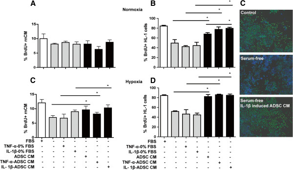
To mimic the behavior of therapeutic cells in the post-infarct cardiac microenvironment, human Adipose Derived Stromal Cells (ADSC) were cultured under hypoxic (2% O2) and pro-inflammatory conditions (IL-1β) for 24h. Serum-free conditioned medium from ADSC primed with hypoxia and/or IL-1β was added to rat neonatal cardiomyocytes and adult cardiomyocytes (HL-1) to assess paracrine-driven changes in cardiomyocyte proliferation rate and induction of myogenic signaling pathways.
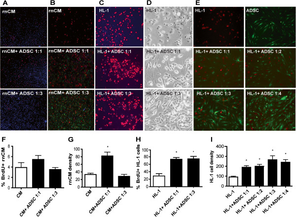
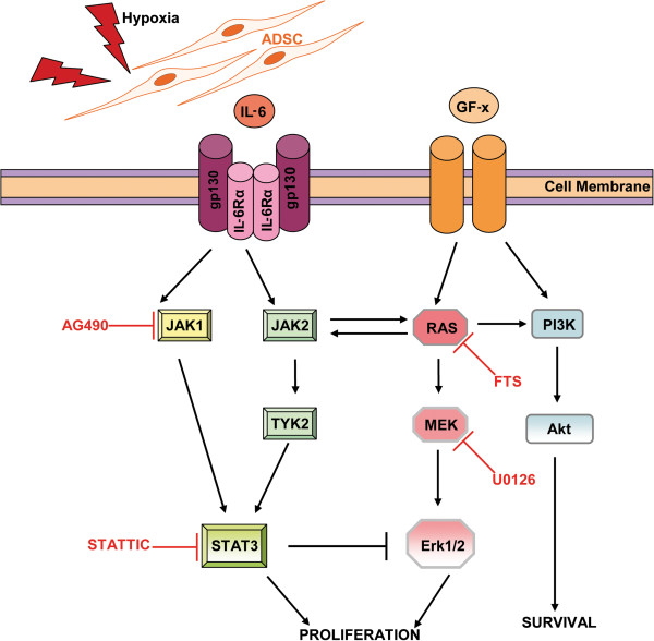
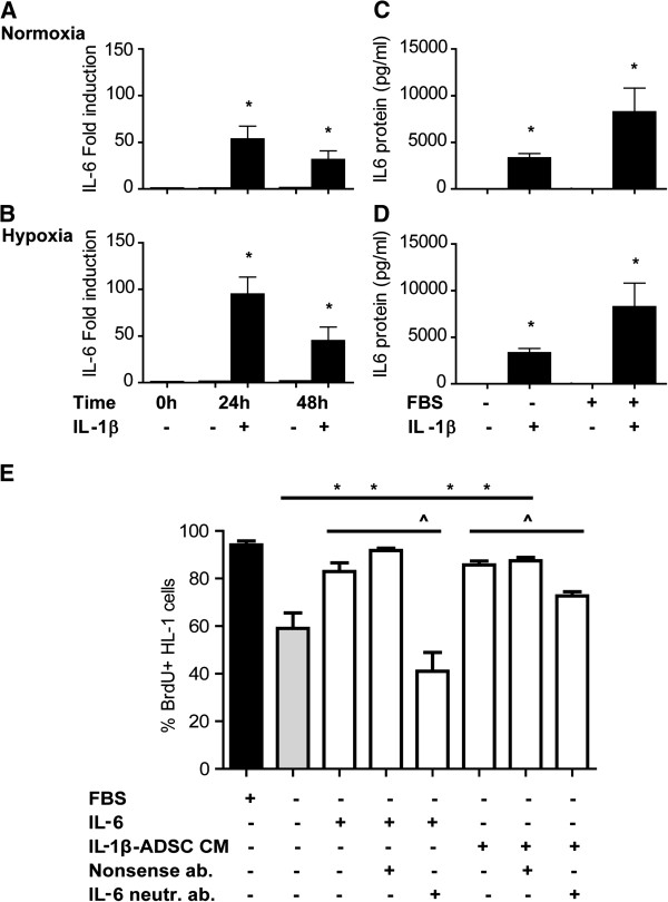
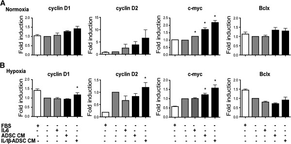
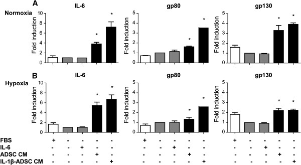
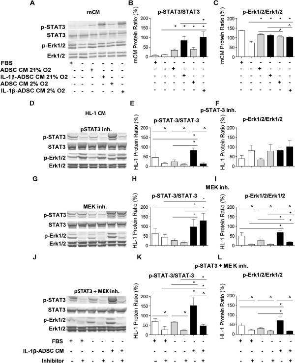
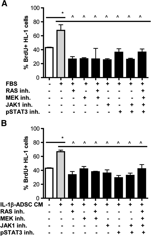
We demonstrate that ADSC enhance the proliferation rate of rat neonatal cardiomyocytes and adult HL-1 cardiomyocytes in a paracrine fashion. ADSC under hypoxia and inflammation in vitro had increased the interleukin-6 (IL-6) gene and protein expression. Similar to conditioned medium of ADSC, treatment of rat neonatal cardiomyocytes and HL-1 with recombinant IL-6 alone also stimulated their proliferation rate. This was corroborated by a strong decrease of cardiomyocyte proliferation after addition of IL-6 neutralizing antibody to conditioned medium of ADSC. The stimulatory effect of ADSC conditioned media or IL-6 was accomplished through activation of both Janus Kinase-Signal Transducer and Activator of Transcription (JAK/STAT) and Mitogen-Activated Protein (MAP) kinases (MAPK) mitogenic signaling pathways.
ADSC are promising therapeutic cells for cardiac stem cell therapy. The inflammatory and hypoxic host post-MI microenvironment enhances the regenerative potential of ADSC to promote the proliferation rate of cardiomyocytes. This was achieved in paracrine manner, which warrants the development of ADSC conditioned medium as an "of-the-shelf" product for treatment of post-myocardial infarction complications.
实验性临床干细胞疗法已被用于缓解急性心肌梗死(AMI)后的不良后果超过十年。梗死心肌的微环境的特点是心肌细胞死亡,这是由缺血和炎症引起的。这些情况可能会对给予的干细胞产生负面影响。由于出生后的心肌细胞增殖率较差,而诱导增殖似乎更为罕见。因此,AMI 后刺激其增殖率至关重要。在化生疾病中,促炎细胞因子白细胞介素 6(IL-6)已被确定为增殖率的有效介质。我们假设 IL-6 可以增加(慢)分裂心肌细胞的增殖率。
为了模拟治疗细胞在梗死心肌微环境中的行为,将人脂肪来源的基质细胞(ADSC)在低氧(2%O2)和促炎条件(IL-1β)下培养 24 小时。用低氧和/或 IL-1β 预培养的 ADSC 的无血清条件培养基被添加到大鼠新生心肌细胞和成年心肌细胞(HL-1)中,以评估旁分泌驱动的心肌细胞增殖率变化和诱导的肌生成信号通路。
我们证明 ADSC 以旁分泌方式增强大鼠新生心肌细胞和成年 HL-1 心肌细胞的增殖率。体外低氧和炎症下的 ADSC 增加了白细胞介素 6(IL-6)基因和蛋白表达。与 ADSC 的条件培养基类似,单独用重组 IL-6 处理大鼠新生心肌细胞和 HL-1 也刺激了它们的增殖率。向 ADSC 的条件培养基中添加 IL-6 中和抗体后,心肌细胞增殖明显减少,证实了这一点。ADSC 条件培养基或 IL-6 的刺激作用是通过激活 Janus 激酶信号转导物和转录激活物(JAK/STAT)和丝裂原激活蛋白(MAP)激酶(MAPK)有丝分裂信号通路来实现的。
ADSC 是心脏干细胞治疗有前途的治疗细胞。AMI 后炎症和低氧的宿主微环境增强了 ADSC 的再生潜力,以促进心肌细胞的增殖率。这是通过旁分泌方式实现的,这需要开发 ADSC 条件培养基作为治疗心肌梗死后并发症的“现成”产品。