• 文献检索
  • 文档翻译
  • 深度研究
  • 学术资讯
  • Suppr Zotero 插件Zotero 插件
  • 邀请有礼
  • 套餐&价格
  • 历史记录
应用&插件
Suppr Zotero 插件Zotero 插件浏览器插件Mac 客户端Windows 客户端微信小程序
定价
高级版会员购买积分包购买API积分包
服务
文献检索文档翻译深度研究API 文档MCP 服务
关于我们
关于 Suppr公司介绍联系我们用户协议隐私条款
关注我们

Suppr 超能文献

核心技术专利:CN118964589B侵权必究
粤ICP备2023148730 号-1Suppr @ 2026

文献检索

告别复杂PubMed语法,用中文像聊天一样搜索,搜遍4000万医学文献。AI智能推荐,让科研检索更轻松。

立即免费搜索

文件翻译

保留排版,准确专业,支持PDF/Word/PPT等文件格式,支持 12+语言互译。

免费翻译文档

深度研究

AI帮你快速写综述,25分钟生成高质量综述,智能提取关键信息,辅助科研写作。

立即免费体验

非成像光驱动功能在常染色体显性视神经萎缩的小鼠模型中得到保留。

Non-image-forming light driven functions are preserved in a mouse model of autosomal dominant optic atrophy.

机构信息

Nuffield Department of Clinical Neurosciences, Nuffield Laboratory of Ophthalmology, University of Oxford, Oxford, United Kingdom.

出版信息

PLoS One. 2013;8(2):e56350. doi: 10.1371/journal.pone.0056350. Epub 2013 Feb 11.

DOI:10.1371/journal.pone.0056350
PMID:23409176
原文链接:https://pmc.ncbi.nlm.nih.gov/articles/PMC3569441/
Abstract

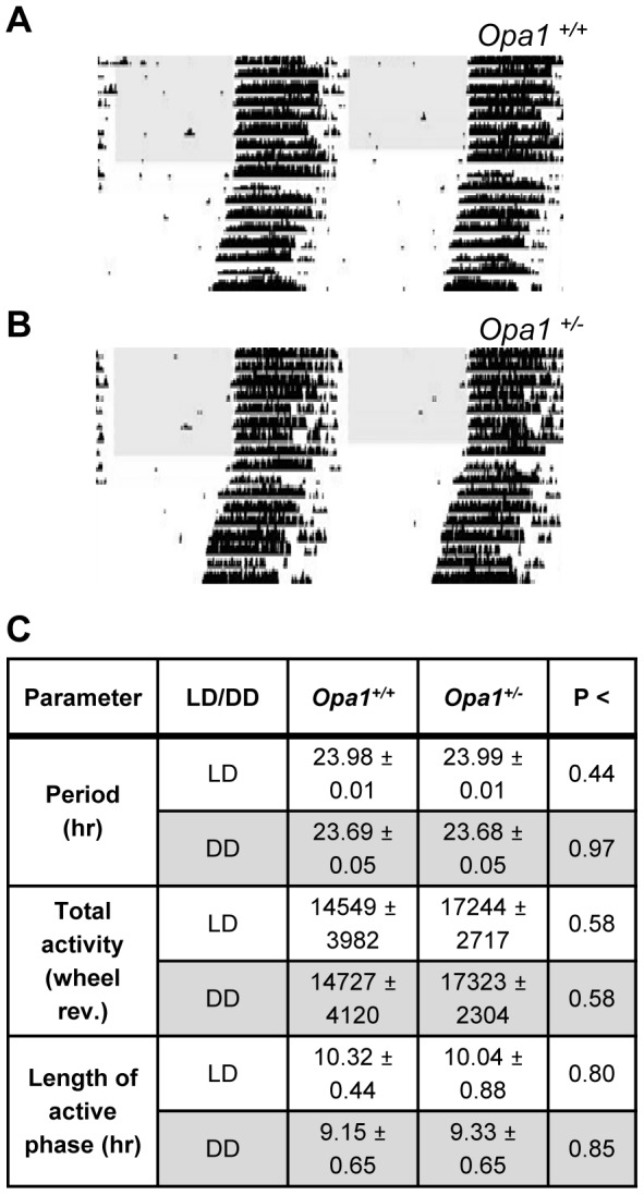
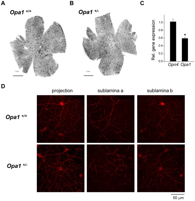
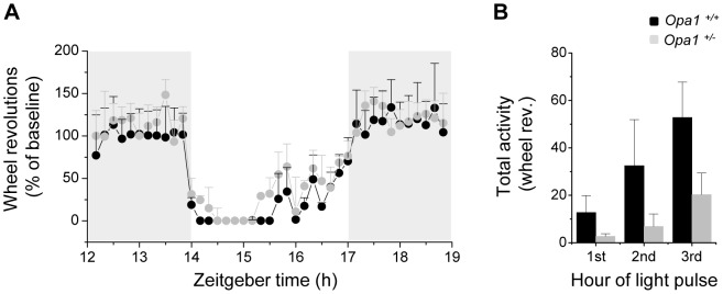
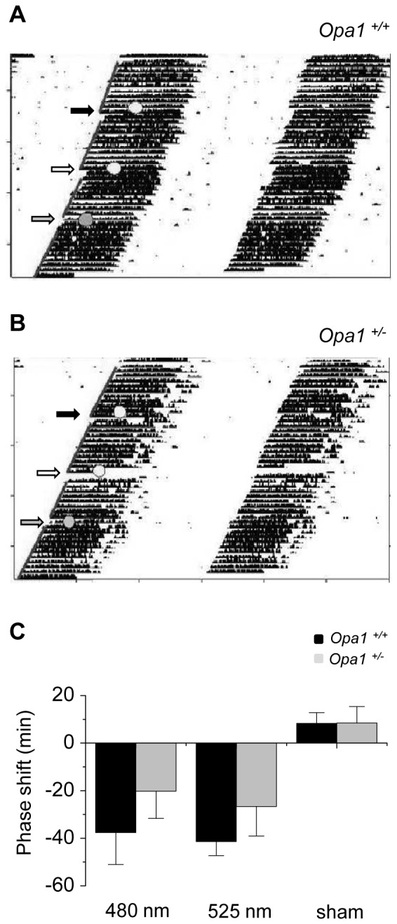
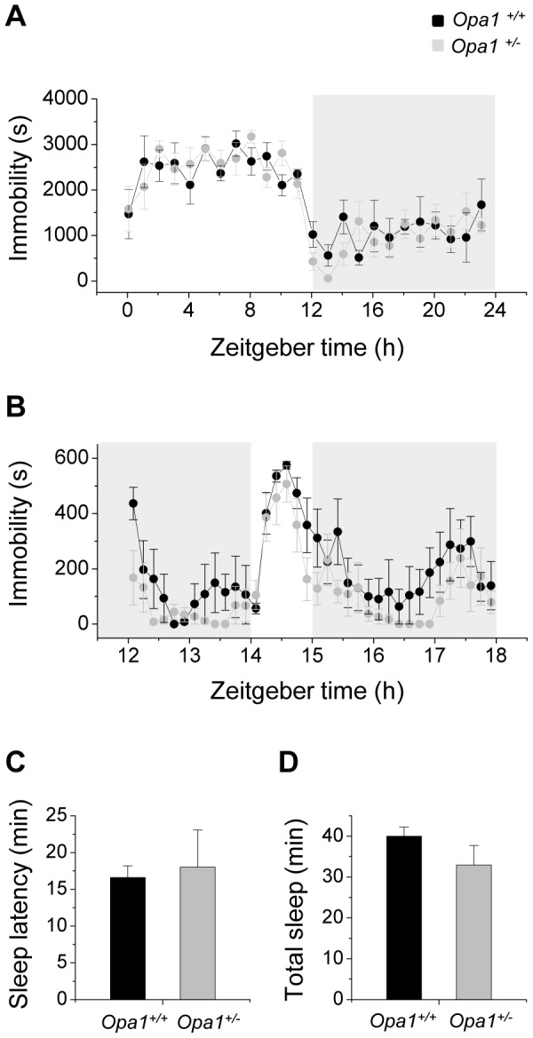
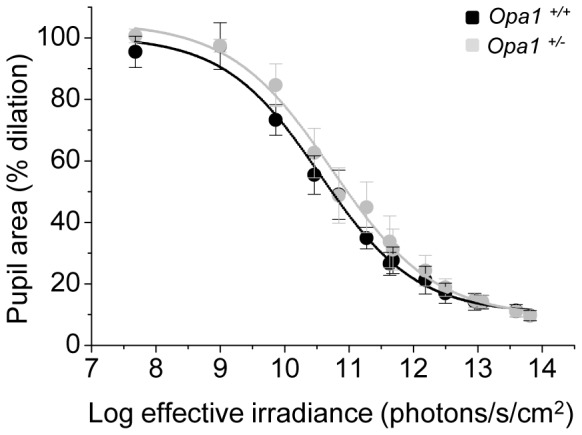
Autosomal dominant optic atrophy (ADOA) is a slowly progressive optic neuropathy that has been associated with mutations of the OPA1 gene. In patients, the disease primarily affects the retinal ganglion cells (RGCs) and causes optic nerve atrophy and visual loss. A subset of RGCs are intrinsically photosensitive, express the photopigment melanopsin and drive non-image-forming (NIF) visual functions including light driven circadian and sleep behaviours and the pupil light reflex. Given the RGC pathology in ADOA, disruption of NIF functions might be predicted. Interestingly in ADOA patients the pupil light reflex was preserved, although NIF behavioural outputs were not examined. The B6; C3-Opa1(Q285STOP) mouse model of ADOA displays optic nerve abnormalities, RGC dendropathy and functional visual disruption. We performed a comprehensive assessment of light driven NIF functions in this mouse model using wheel running activity monitoring, videotracking and pupillometry. Opa1 mutant mice entrained their activity rhythm to the external light/dark cycle, suppressed their activity in response to acute light exposure at night, generated circadian phase shift responses to 480 nm and 525 nm pulses, demonstrated immobility-defined sleep induction following exposure to a brief light pulse at night and exhibited an intensity dependent pupil light reflex. There were no significant differences in any parameter tested relative to wildtype littermate controls. Furthermore, there was no significant difference in the number of melanopsin-expressing RGCs, cell morphology or melanopsin transcript levels between genotypes. Taken together, these findings suggest the preservation of NIF functions in Opa1 mutants. The results provide support to growing evidence that the melanopsin-expressing RGCs are protected in mitochondrial optic neuropathies.

摘要

常染色体显性视神经萎缩(ADOA)是一种进行性缓慢的视神经病变,与 OPA1 基因突变有关。在患者中,该疾病主要影响视网膜神经节细胞(RGC),导致视神经萎缩和视力丧失。RGC 中有一部分是内在光敏的,表达感光色素黑视蛋白,并驱动非成像(NIF)视觉功能,包括光驱动的昼夜节律和睡眠行为以及瞳孔光反射。鉴于 ADOA 中的 RGC 病理学,预计会破坏 NIF 功能。有趣的是,在 ADOA 患者中,瞳孔光反射得以保留,尽管没有检查 NIF 行为输出。B6; C3-Opa1(Q285STOP)ADOA 小鼠模型显示视神经异常、RGC 树突病和功能视觉障碍。我们使用轮式跑步活动监测、视频跟踪和瞳孔计对该小鼠模型中的光驱动 NIF 功能进行了全面评估。Opa1 突变小鼠使它们的活动节律与外部明暗周期同步,在夜间急性光照暴露时抑制其活动,对 480nm 和 525nm 脉冲产生昼夜节律相位移动反应,在夜间短暂光脉冲暴露后表现出不动定义的睡眠诱导,并表现出强度依赖性瞳孔光反射。与野生型同窝对照相比,在任何测试参数上均无显著差异。此外,在基因型之间,黑视蛋白表达的 RGC 数量、细胞形态或黑视蛋白转录水平均无显著差异。综上所述,这些发现表明 Opa1 突变体中 NIF 功能得以保留。这些结果为越来越多的证据提供了支持,即表达黑视蛋白的 RGC 在线粒体视神经病变中受到保护。

https://cdn.ncbi.nlm.nih.gov/pmc/blobs/29e4/3569441/faf724ce5fa0/pone.0056350.g006.jpg
https://cdn.ncbi.nlm.nih.gov/pmc/blobs/29e4/3569441/af59e826bbc2/pone.0056350.g001.jpg
https://cdn.ncbi.nlm.nih.gov/pmc/blobs/29e4/3569441/cd2ab4a626dd/pone.0056350.g002.jpg
https://cdn.ncbi.nlm.nih.gov/pmc/blobs/29e4/3569441/cc445bda58b6/pone.0056350.g003.jpg
https://cdn.ncbi.nlm.nih.gov/pmc/blobs/29e4/3569441/6a9eb112869e/pone.0056350.g004.jpg
https://cdn.ncbi.nlm.nih.gov/pmc/blobs/29e4/3569441/b28ad2117205/pone.0056350.g005.jpg
https://cdn.ncbi.nlm.nih.gov/pmc/blobs/29e4/3569441/faf724ce5fa0/pone.0056350.g006.jpg

相似文献

1
Non-image-forming light driven functions are preserved in a mouse model of autosomal dominant optic atrophy.非成像光驱动功能在常染色体显性视神经萎缩的小鼠模型中得到保留。
PLoS One. 2013;8(2):e56350. doi: 10.1371/journal.pone.0056350. Epub 2013 Feb 11.
2
Influence of Opa1 Mutation on Survival and Function of Retinal Ganglion Cells.Opa1突变对视网膜神经节细胞存活和功能的影响。
Invest Ophthalmol Vis Sci. 2015 Jul;56(8):4835-45. doi: 10.1167/iovs.15-16743.
3
Specific deficits in visual electrophysiology in a mouse model of dominant optic atrophy.在显性视神经萎缩的小鼠模型中存在视觉电生理学的特定缺陷。
Exp Eye Res. 2011 Nov;93(5):771-7. doi: 10.1016/j.exer.2011.07.004. Epub 2011 Jul 22.
4
Opa1 deficiency in a mouse model of autosomal dominant optic atrophy impairs mitochondrial morphology, optic nerve structure and visual function.常染色体显性视神经萎缩小鼠模型中的Opa1缺陷会损害线粒体形态、视神经结构和视觉功能。
Hum Mol Genet. 2007 Jun 1;16(11):1307-18. doi: 10.1093/hmg/ddm079. Epub 2007 Apr 11.
5
Melanopsin retinal ganglion cells are resistant to neurodegeneration in mitochondrial optic neuropathies.黑视蛋白视网膜神经节细胞对线粒体视神经病变的神经退行性变有抗性。
Brain. 2010 Aug;133(Pt 8):2426-38. doi: 10.1093/brain/awq155. Epub 2010 Jul 21.
6
Red Light Irradiation In Vivo Upregulates DJ-1 in the Retinal Ganglion Cell Layer and Protects against Axotomy-Related Dendritic Pruning.红光体内辐照上调视网膜神经节细胞层中的 DJ-1 并保护其免受轴突切断相关树突修剪。
Int J Mol Sci. 2021 Aug 4;22(16):8380. doi: 10.3390/ijms22168380.
7
OPA1 deficiency associated with increased autophagy in retinal ganglion cells in a murine model of dominant optic atrophy.在显性遗传性视神经萎缩小鼠模型中,OPA1缺乏与视网膜神经节细胞自噬增加有关。
Invest Ophthalmol Vis Sci. 2009 Jun;50(6):2567-71. doi: 10.1167/iovs.08-2913. Epub 2009 Feb 21.
8
Opa1 deficiency in a mouse model of dominant optic atrophy leads to retinal ganglion cell dendropathy.在显性视神经萎缩的小鼠模型中,opa1 缺失导致视网膜神经节细胞树突病变。
Brain. 2010 Oct;133(10):2942-51. doi: 10.1093/brain/awq218. Epub 2010 Sep 3.
9
A randomized, placebo-controlled trial of the benzoquinone idebenone in a mouse model of OPA1-related dominant optic atrophy reveals a limited therapeutic effect on retinal ganglion cell dendropathy and visual function.在OPA1相关显性遗传性视神经萎缩小鼠模型中进行的关于苯醌艾地苯醌的随机、安慰剂对照试验显示,其对视网膜神经节细胞树突病变和视觉功能的治疗效果有限。
Neuroscience. 2016 Apr 5;319:92-106. doi: 10.1016/j.neuroscience.2016.01.042. Epub 2016 Jan 25.
10
Electrophysiological and histologic assessment of retinal ganglion cell fate in a mouse model for OPA1-associated autosomal dominant optic atrophy.OPA1 相关常染色体显性视神经萎缩小鼠模型中视网膜神经节细胞命运的电生理学和组织学评估。
Invest Ophthalmol Vis Sci. 2010 Mar;51(3):1424-31. doi: 10.1167/iovs.09-3606. Epub 2009 Oct 15.

引用本文的文献

1
Retinal ganglion cell repopulation for vision restoration in optic neuropathy: a roadmap from the RReSTORe Consortium.视网膜神经节细胞再灌注以恢复视神经病变的视力:RReSTORe 联盟的路线图。
Mol Neurodegener. 2023 Sep 21;18(1):64. doi: 10.1186/s13024-023-00655-y.
2
Phenotype Characterization of a Mice Genetic Model of Absolute Blindness.绝对盲小鼠遗传模型的表型特征分析。
Int J Mol Sci. 2022 Jul 24;23(15):8152. doi: 10.3390/ijms23158152.
3
Differential susceptibility of retinal ganglion cell subtypes against neurodegenerative diseases.

本文引用的文献

1
Form and function of the M4 cell, an intrinsically photosensitive retinal ganglion cell type contributing to geniculocortical vision.M4 细胞的形态和功能,作为一种内在光敏感的视网膜神经节细胞类型,有助于视束-皮质视觉。
J Neurosci. 2012 Sep 26;32(39):13608-20. doi: 10.1523/JNEUROSCI.1422-12.2012.
2
Rapid assessment of sleep-wake behavior in mice.快速评估小鼠的睡眠-觉醒行为。
J Biol Rhythms. 2012 Feb;27(1):48-58. doi: 10.1177/0748730411431550.
3
Specific deficits in visual electrophysiology in a mouse model of dominant optic atrophy.
视网膜神经节细胞亚型对神经退行性疾病的易感性差异。
Graefes Arch Clin Exp Ophthalmol. 2022 Jun;260(6):1807-1821. doi: 10.1007/s00417-022-05556-2. Epub 2022 Jan 17.
4
Dominant Optic Atrophy (DOA): Modeling the Kaleidoscopic Roles of OPA1 in Mitochondrial Homeostasis.显性视神经萎缩(DOA):模拟OPA1在线粒体稳态中的多种作用
Front Neurol. 2021 Jun 9;12:681326. doi: 10.3389/fneur.2021.681326. eCollection 2021.
5
Retinal Ganglion Cells-Diversity of Cell Types and Clinical Relevance.视网膜神经节细胞——细胞类型的多样性及临床相关性
Front Neurol. 2021 May 21;12:661938. doi: 10.3389/fneur.2021.661938. eCollection 2021.
6
Opa1 Deficiency Leads to Diminished Mitochondrial Bioenergetics With Compensatory Increased Mitochondrial Motility.OPA1 缺失导致线粒体生物能量学降低,同时伴有代偿性的线粒体运动性增加。
Invest Ophthalmol Vis Sci. 2020 Jun 3;61(6):42. doi: 10.1167/iovs.61.6.42.
7
Melanopsin Retinal Ganglion Cells and Pupil: Clinical Implications for Neuro-Ophthalmology.黑视蛋白视网膜神经节细胞与瞳孔:对神经眼科的临床意义
Front Neurol. 2018 Dec 7;9:1047. doi: 10.3389/fneur.2018.01047. eCollection 2018.
8
The Pupillary Light Reflex in Idiopathic Intracranial Hypertension.特发性颅内高压中的瞳孔光反射
Invest Ophthalmol Vis Sci. 2016 Jan 1;57(1):23-9. doi: 10.1167/iovs.15-18181.
9
Comparison of acute non-visual bright light responses in patients with optic nerve disease, glaucoma and healthy controls.视神经疾病、青光眼患者与健康对照者急性非视觉强光反应的比较。
Sci Rep. 2015 Oct 19;5:15185. doi: 10.1038/srep15185.
10
Differential monocular vs. binocular pupil responses from melanopsin-based photoreception in patients with anterior ischemic optic neuropathy.前部缺血性视神经病变患者基于黑视蛋白的光感受器产生的单眼与双眼瞳孔反应差异
Sci Rep. 2015 Jun 15;5:10780. doi: 10.1038/srep10780.
在显性视神经萎缩的小鼠模型中存在视觉电生理学的特定缺陷。
Exp Eye Res. 2011 Nov;93(5):771-7. doi: 10.1016/j.exer.2011.07.004. Epub 2011 Jul 22.
4
Photoentrainment and pupillary light reflex are mediated by distinct populations of ipRGCs.光生物周期节律与瞳孔对光反射由不同的 ipRGC 群体介导。
Nature. 2011 Jul 17;476(7358):92-5. doi: 10.1038/nature10206.
5
The circadian response of intrinsically photosensitive retinal ganglion cells.视网膜神经节细胞的昼夜节律反应。
PLoS One. 2011 Mar 14;6(3):e17860. doi: 10.1371/journal.pone.0017860.
6
Opa1 deficiency in a mouse model of dominant optic atrophy leads to retinal ganglion cell dendropathy.在显性视神经萎缩的小鼠模型中,opa1 缺失导致视网膜神经节细胞树突病变。
Brain. 2010 Oct;133(10):2942-51. doi: 10.1093/brain/awq218. Epub 2010 Sep 3.
7
Melanopsin retinal ganglion cells are resistant to neurodegeneration in mitochondrial optic neuropathies.黑视蛋白视网膜神经节细胞对线粒体视神经病变的神经退行性变有抗性。
Brain. 2010 Aug;133(Pt 8):2426-38. doi: 10.1093/brain/awq155. Epub 2010 Jul 21.
8
Melanopsin-expressing retinal ganglion-cell photoreceptors: cellular diversity and role in pattern vision.表达黑视蛋白的视网膜神经节细胞光感受器:细胞多样性及其在模式视觉中的作用。
Neuron. 2010 Jul 15;67(1):49-60. doi: 10.1016/j.neuron.2010.05.023.
9
Suppression of rat retinal ganglion cell death by PACAP following transient ischemia induced by high intraocular pressure.高眼压诱导短暂性缺血后 PACAP 对大鼠视网膜神经节细胞死亡的抑制作用。
J Mol Neurosci. 2011 Jan;43(1):30-4. doi: 10.1007/s12031-010-9410-5. Epub 2010 Jun 29.
10
Selective wavelength pupillometry in Leber hereditary optic neuropathy.Leber遗传性视神经病变中的选择性波长瞳孔测量法。
Clin Exp Ophthalmol. 2010 Apr;38(3):322-4. doi: 10.1111/j.1442-9071.2010.02212.x.