School of Life, Sport and Social Sciences, Edinburgh Napier University, Edinburgh, United Kingdom.
PLoS One. 2013;8(2):e56263. doi: 10.1371/journal.pone.0056263. Epub 2013 Feb 15.
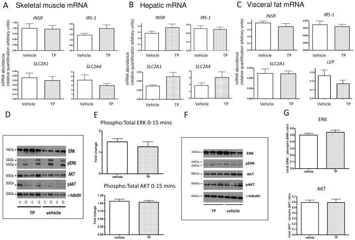
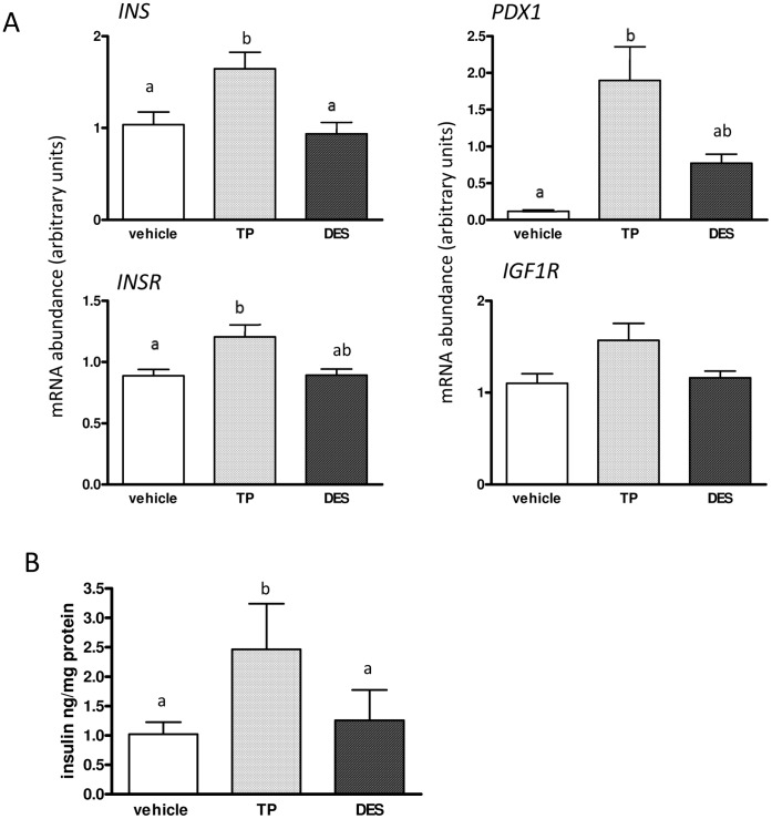
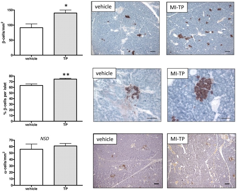
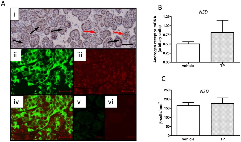
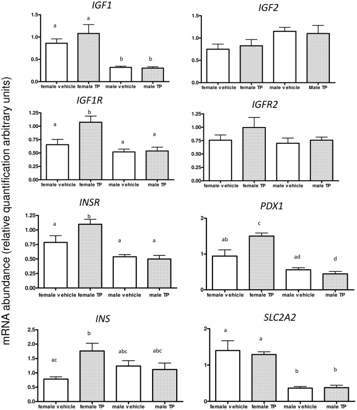
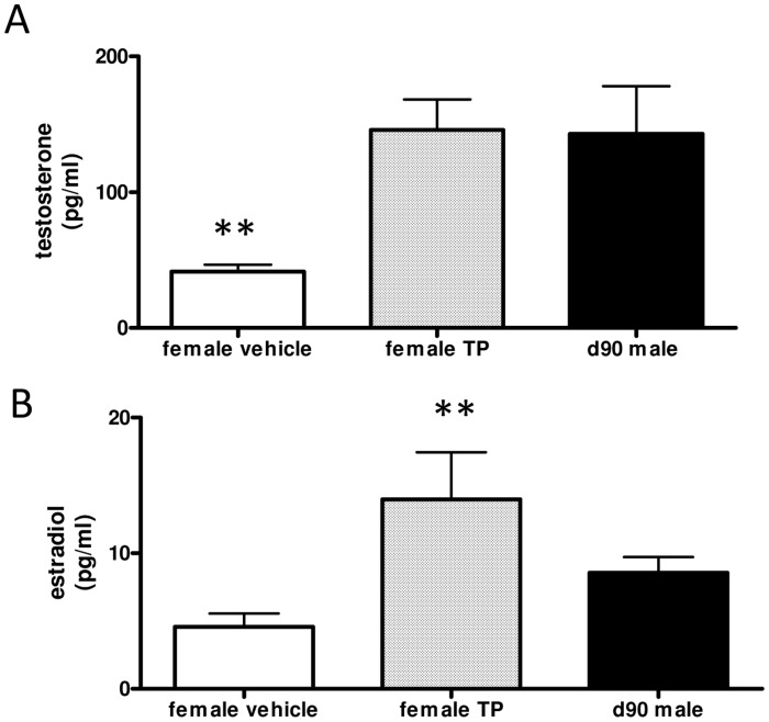
Using an ovine model of polycystic ovary syndrome (PCOS), (pregnant ewes injected with testosterone propionate (TP) (100 mg twice weekly) from day (d)62 to d102 of d147 gestation (maternal injection - MI-TP)), we previously reported female offspring with normal glucose tolerance but hyperinsulinemia. We therefore examined insulin signalling and pancreatic morphology in these offspring using quantitative (Q) RT-PCR and western blotting. In addition the fetal pancreatic responses to MI-TP, and androgenic and estrogenic contributions to such responses (direct fetal injection (FI) of TP (20 mg) or diethylstilbestrol (DES) (20 mg) at d62 and d82 gestation) were assessed at d90 gestation. Fetal plasma was assayed for insulin, testosterone and estradiol, pancreatic tissue was cultured, and expression of key β-cell developmental genes was assessed by QRT-PCR. In female d62MI-TP offspring insulin signalling was unaltered but there was a pancreatic phenotype with increased numbers of β-cells (P<0.05). The fetal pancreas expressed androgen receptors in islets and genes involved in β-cell development and function (PDX1, IGF1R, INSR and INS) were up-regulated in female fetuses after d62MI-TP treatment (P<0.05-0.01). In addition the d62MI-TP pancreas showed increased insulin secretion under euglycaemic conditions (P<0.05) in vitro. The same effects were not seen in the male fetal pancreas or when MI-TP was started at d30, before the male programming window. As d62MI-TP increased both fetal plasma testosterone (P<0.05) and estradiol concentrations (P<0.05) we assessed the relative contribution of androgens and estrogens. FI-TP (commencing d62) (not FI-DES treatment) caused elevated basal insulin secretion in vitro and the genes altered by d62MI-TP treatment were similarly altered by FI-TP but not FI-DES. In conclusion, androgen over-exposure alters fetal pancreatic development and β-cell numbers in offspring. These data suggest that that there may be a primary pancreatic phenotype in models of PCOS, and that there may be a distinct male and female pancreas.
我们使用多囊卵巢综合征(PCOS)的绵羊模型(从妊娠第 147 天(d)62 至 d102 每天两次给怀孕母羊注射 100mg 丙酸睾酮(TP)(母体注射-MI-TP)),此前报道过葡萄糖耐量正常但胰岛素血症的雌性后代。因此,我们使用定量(Q)RT-PCR 和 Western blot 检查了这些后代的胰岛素信号和胰腺形态。此外,我们还评估了 MI-TP 对胎儿胰腺的作用,以及雄激素和雌激素对这种作用的贡献(在妊娠第 62 天和第 82 天直接向胎儿注射 20mgTP 或己烯雌酚(DES))。在妊娠第 90 天,检测胎儿血浆胰岛素、睾酮和雌二醇,培养胰腺组织,并通过 QRT-PCR 评估关键β细胞发育基因的表达。在雌性 d62MI-TP 后代中,胰岛素信号未改变,但胰腺表型显示β细胞数量增加(P<0.05)。胎儿胰岛中表达雄激素受体,β细胞发育和功能相关基因(PDX1、IGF1R、INSR 和 INS)在 MI-TP 处理后(P<0.05-0.01)雌性胎儿中上调。此外,在体外,d62MI-TP 胰腺在正常血糖条件下显示出增加的胰岛素分泌(P<0.05)。在雄性胎儿胰腺或在雄性编程窗口之前(在第 30 天开始)开始 MI-TP 治疗时,没有观察到相同的效果。由于 d62MI-TP 增加了胎儿血浆睾酮(P<0.05)和雌二醇浓度(P<0.05),我们评估了雄激素和雌激素的相对贡献。FI-TP(从第 62 天开始)(而不是 FI-DES 治疗)导致体外基础胰岛素分泌增加,d62MI-TP 治疗改变的基因也被 FI-TP 改变,但不是 FI-DES。总之,雄激素过度暴露改变了后代胎儿胰腺发育和β细胞数量。这些数据表明,在 PCOS 模型中可能存在原发性胰腺表型,并且可能存在明显的雄性和雌性胰腺。