Department of Orthopaedic Surgery, School of Medicine, Fujita Health University, Toyoake, Aichi, Japan.
PLoS One. 2013;8(2):e56641. doi: 10.1371/journal.pone.0056641. Epub 2013 Feb 14.
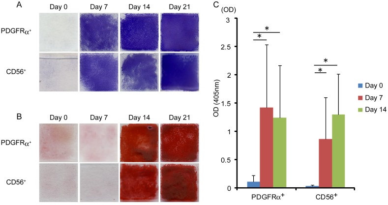
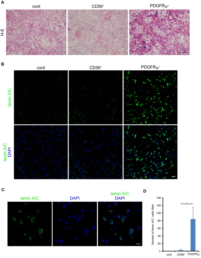
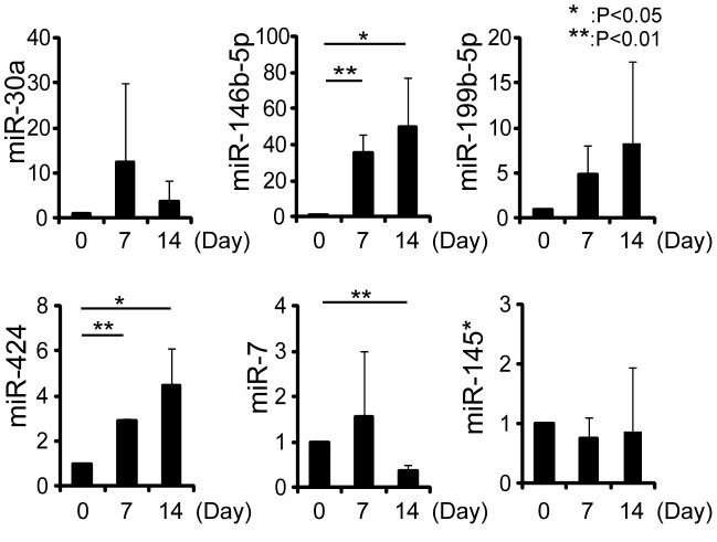
Heterotopic ossification (HO) is defined as the formation of ectopic bone in soft tissue outside the skeletal tissue. HO is thought to result from aberrant differentiation of osteogenic progenitors within skeletal muscle. However, the precise origin of HO is still unclear. Skeletal muscle contains two kinds of progenitor cells, myogenic progenitors and mesenchymal progenitors. Myogenic and mesenchymal progenitors in human skeletal muscle can be identified as CD56(+) and PDGFRα(+) cells, respectively. The purpose of this study was to investigate the osteogenic differentiation potential of human skeletal muscle-derived progenitors. Both CD56(+) cells and PDGFRα(+) cells showed comparable osteogenic differentiation potential in vitro. However, in an in vivo ectopic bone formation model, PDGFRα(+) cells formed bone-like tissue and showed successful engraftment, while CD56(+) cells did not form bone-like tissue and did not adapt to an osteogenic environment. Immunohistological analysis of human HO sample revealed that many PDGFRα(+) cells were localized in proximity to ectopic bone formed in skeletal muscle. MicroRNAs (miRNAs) are known to regulate many biological processes including osteogenic differentiation. We investigated the participation of miRNAs in the osteogenic differentiation of PDGFRα(+) cells by using microarray. We identified miRNAs that had not been known to be involved in osteogenesis but showed dramatic changes during osteogenic differentiation of PDGFRα(+) cells. Upregulation of miR-146b-5p and -424 and downregulation of miR-7 during osteogenic differentiation of PDGFRα(+) cells were confirmed by quantitative real-time RT-PCR. Inhibition of upregulated miRNAs, miR-146b-5p and -424, resulted in the suppression of osteocyte maturation, suggesting that these two miRNAs have the positive role in the osteogenesis of PDGFRα(+) cells. Our results suggest that PDGFRα(+) cells may be the major source of HO and that the newly identified miRNAs may regulate osteogenic differentiation process of PDGFRα(+) cells.
异位骨化(HO)被定义为在骨骼组织外的软组织中形成异位骨。HO 被认为是骨骼肌肉内成骨祖细胞异常分化的结果。然而,HO 的确切起源仍不清楚。骨骼肌包含两种祖细胞,即肌源性祖细胞和间充质祖细胞。人骨骼肌中的肌源性和间充质祖细胞分别可以被鉴定为 CD56(+)和 PDGFRα(+)细胞。本研究旨在探讨人骨骼肌源性祖细胞的成骨分化潜能。CD56(+)细胞和 PDGFRα(+)细胞在体外均具有相当的成骨分化潜能。然而,在体内异位骨形成模型中,PDGFRα(+)细胞形成骨样组织并成功植入,而 CD56(+)细胞则未形成骨样组织,也未适应成骨环境。对人 HO 样本的免疫组织化学分析表明,许多 PDGFRα(+)细胞定位于骨骼肌中形成的异位骨附近。已知 microRNAs(miRNAs)调节包括成骨分化在内的许多生物学过程。我们通过微阵列研究了 miRNAs 在 PDGFRα(+)细胞成骨分化中的参与情况。我们鉴定了一些以前不参与成骨但在 PDGFRα(+)细胞成骨分化过程中表现出明显变化的 miRNAs。定量实时 RT-PCR 证实 PDGFRα(+)细胞成骨分化过程中 miR-146b-5p 和 -424 的上调和 miR-7 的下调。上调的 miRNAs,miR-146b-5p 和 -424 的抑制导致成骨细胞成熟受到抑制,表明这两个 miRNA 在 PDGFRα(+)细胞的成骨中具有积极作用。我们的研究结果表明,PDGFRα(+)细胞可能是 HO 的主要来源,而新鉴定的 miRNAs 可能调节 PDGFRα(+)细胞的成骨分化过程。