Zhang Zhongyu, Zhang Yan, Xiao Huizhen, Liang Xiaolei, Sun Dawei, Peng Shaomin
Department of Ophthalmology, the Second Affiliated Hospital of Harbin Medical University, Harbin, China.
Mol Vis. 2012;18:2961-75. Epub 2012 Dec 15.
The molecular mechanisms associated with human retinal pigment epithelium (RPE) development constitute the basis for cell replacement therapy for the treatment of retinal degenerative diseases. In the current study, gene expression analysis of the human fetal RPE during development was performed and was compared with the human native RPE.
Microdissection of the human RPE at three time points (13 weeks and 16 weeks of gestation and in mature adult eyes) was performed, and total RNA was isolated. Equal amounts of RNA were pooled from two or three independent donor eyes for each time point in each group. Gene expression was analyzed by hybridization to microarray chips. Validation was accomplished by comparing the microarray expression profiles with quantitative real-time reverse transcriptase-polymerase chain reaction (qRT(2)-PCR) analysis of selected genes and by comparing selected expression profiles with predicted profiles based on previous studies.
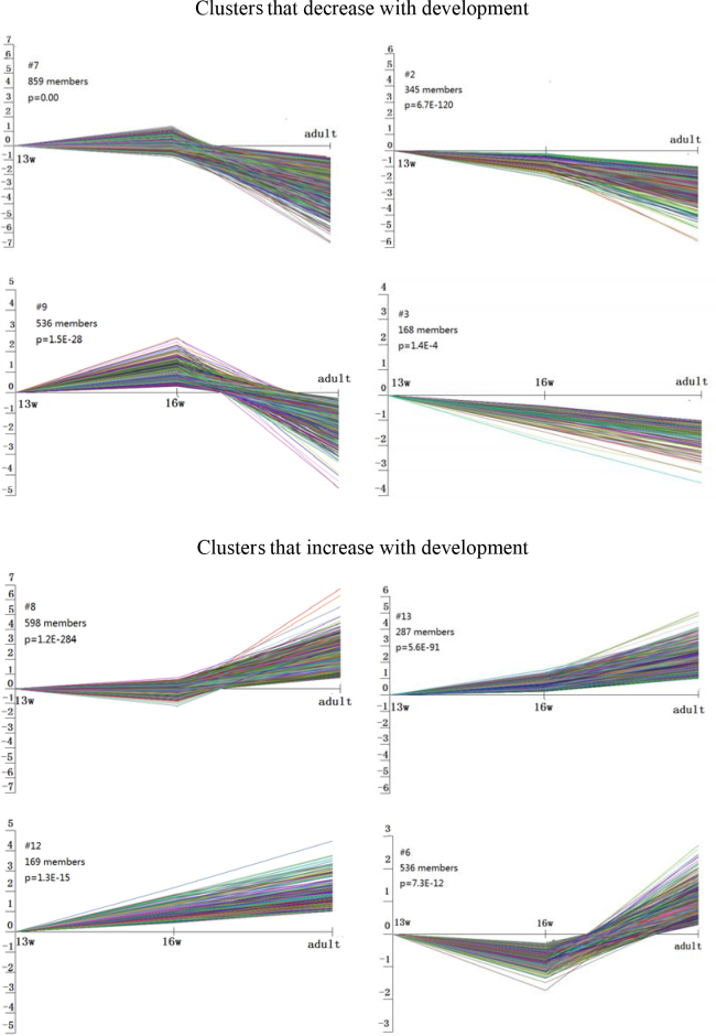
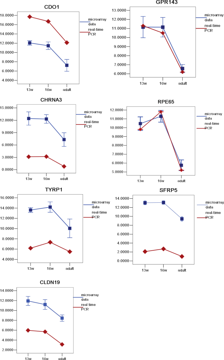
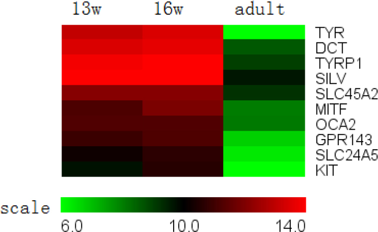
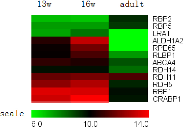
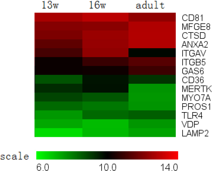
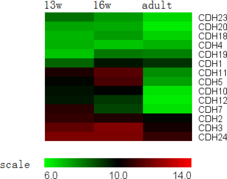
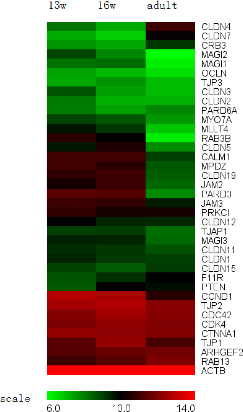
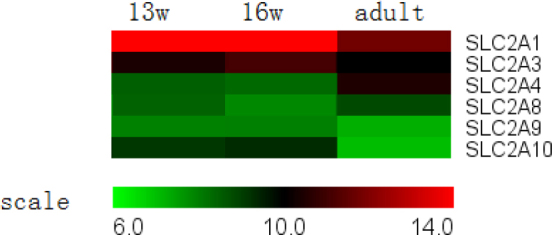
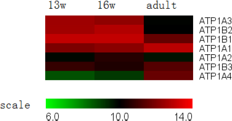
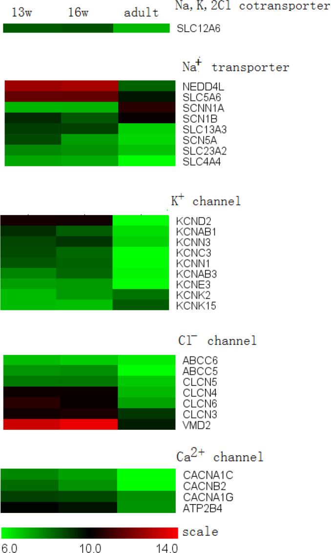
Of the 45,033 probe sets on the microarray, 30,736 were detected. A total of 3,498 differentially expressed genes could be clustered into eight patterns of expression that were statistically significant. Analysis of the expression patterns of genes coding for key functions (pigment synthesis, visual cycle, phagocytosis, adherens and tight junctions, and transcellular transport) indicated that the human RPE achieves a high degree of maturity during early pregnancy. Compared with 154 signature genes in the RPE, 148 candidate genes were identified in this study, including 53 downregulated genes and 5 upregulated genes. The qRT(2)-PCR results showed similar expression trends to those obtained by microarray analysis at the three time points.
This study demonstrated gene expression profiles in the human RPE during normal development. These findings indicate that the human RPE has different expression patterns than those of other animals. The results of this study may be helpful in furthering the understanding of the developmental processes occurring in humans and of the differentiation of RPE cells derived from human embryonic stem cells and from human induced pluripotent stem cells.
与人类视网膜色素上皮(RPE)发育相关的分子机制构成了用于治疗视网膜退行性疾病的细胞替代疗法的基础。在本研究中,对人类胎儿发育过程中的RPE进行了基因表达分析,并与人类天然RPE进行了比较。
在三个时间点(妊娠13周和16周以及成年成熟眼睛)对人类RPE进行显微切割,并分离总RNA。每组中每个时间点从两到三只独立供体眼睛中收集等量的RNA。通过与微阵列芯片杂交分析基因表达。通过将微阵列表达谱与所选基因的定量实时逆转录聚合酶链反应(qRT(2)-PCR)分析进行比较,并将所选表达谱与基于先前研究的预测谱进行比较来完成验证。
在微阵列上的45,033个探针集中,检测到30,736个。总共3,498个差异表达基因可聚类为八种具有统计学意义的表达模式。对编码关键功能(色素合成、视觉循环、吞噬作用、黏附连接和紧密连接以及跨细胞运输)的基因的表达模式分析表明,人类RPE在妊娠早期达到高度成熟。与RPE中的154个特征基因相比,本研究鉴定出148个候选基因,包括53个下调基因和5个上调基因。qRT(2)-PCR结果在三个时间点显示出与微阵列分析相似的表达趋势。
本研究展示了人类RPE在正常发育过程中的基因表达谱。这些发现表明人类RPE具有与其他动物不同的表达模式。本研究结果可能有助于进一步了解人类发生的发育过程以及源自人类胚胎干细胞和人类诱导多能干细胞的RPE细胞的分化。