• 文献检索
  • 文档翻译
  • 深度研究
  • 学术资讯
  • Suppr Zotero 插件Zotero 插件
  • 邀请有礼
  • 套餐&价格
  • 历史记录
应用&插件
Suppr Zotero 插件Zotero 插件浏览器插件Mac 客户端Windows 客户端微信小程序
定价
高级版会员购买积分包购买API积分包
服务
文献检索文档翻译深度研究API 文档MCP 服务
关于我们
关于 Suppr公司介绍联系我们用户协议隐私条款
关注我们

Suppr 超能文献

核心技术专利:CN118964589B侵权必究
粤ICP备2023148730 号-1Suppr @ 2026

文献检索

告别复杂PubMed语法,用中文像聊天一样搜索,搜遍4000万医学文献。AI智能推荐,让科研检索更轻松。

立即免费搜索

文件翻译

保留排版,准确专业,支持PDF/Word/PPT等文件格式,支持 12+语言互译。

免费翻译文档

深度研究

AI帮你快速写综述,25分钟生成高质量综述,智能提取关键信息,辅助科研写作。

立即免费体验

应用高阶扩散张量和磁化率张量 MRI 探测脑白质微观结构

Probing white-matter microstructure with higher-order diffusion tensors and susceptibility tensor MRI.

机构信息

Brain Imaging and Analysis Center, School of Medicine, Duke University Durham, NC, USA ; Department of Radiology, Duke University Durham, NC, USA.

出版信息

Front Integr Neurosci. 2013 Mar 6;7:11. doi: 10.3389/fnint.2013.00011. eCollection 2013.

DOI:10.3389/fnint.2013.00011
PMID:23507987
原文链接:https://pmc.ncbi.nlm.nih.gov/articles/PMC3589706/
Abstract

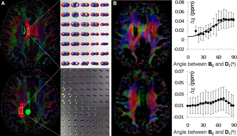
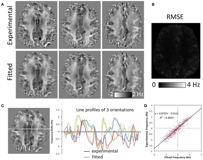
Diffusion MRI has become an invaluable tool for studying white matter microstructure and brain connectivity. The emergence of quantitative susceptibility mapping and susceptibility tensor imaging (STI) has provided another unique tool for assessing the structure of white matter. In the highly ordered white matter structure, diffusion MRI measures hindered water mobility induced by various tissue and cell membranes, while susceptibility sensitizes to the molecular composition and axonal arrangement. Integrating these two methods may produce new insights into the complex physiology of white matter. In this study, we investigated the relationship between diffusion and magnetic susceptibility in the white matter. Experiments were conducted on phantoms and human brains in vivo. Diffusion properties were quantified with the diffusion tensor model and also with the higher order tensor model based on the cumulant expansion. Frequency shift and susceptibility tensor were measured with quantitative susceptibility mapping and susceptibility tensor imaging. These diffusion and susceptibility quantities were compared and correlated in regions of single fiber bundles and regions of multiple fiber orientations. Relationships were established with similarities and differences identified. It is believed that diffusion MRI and susceptibility MRI provide complementary information of the microstructure of white matter. Together, they allow a more complete assessment of healthy and diseased brains.

摘要

扩散 MRI 已成为研究白质微观结构和大脑连接的不可或缺的工具。定量磁化率映射和磁化张量成像(STI)的出现为评估白质结构提供了另一种独特的工具。在高度有序的白质结构中,扩散 MRI 测量由各种组织和细胞膜引起的水流动性受阻,而磁化率对分子组成和轴突排列敏感。整合这两种方法可能会对白质的复杂生理学产生新的认识。在这项研究中,我们研究了白质中扩散和磁化率之间的关系。在体模和人体大脑上进行了实验。使用扩散张量模型和基于累积展开的高阶张量模型来量化扩散特性。使用定量磁化率映射和磁化张量成像测量频率偏移和磁化率张量。在单纤维束区域和多纤维取向区域比较和关联这些扩散和磁化率量。确定了相似性和差异性。据信,扩散 MRI 和磁化率 MRI 提供了白质微观结构的互补信息。它们共同允许对健康和患病的大脑进行更全面的评估。

https://cdn.ncbi.nlm.nih.gov/pmc/blobs/6821/3589706/fa939c2b8306/fnint-07-00011-g0005.jpg
https://cdn.ncbi.nlm.nih.gov/pmc/blobs/6821/3589706/6cbf37579057/fnint-07-00011-g0001.jpg
https://cdn.ncbi.nlm.nih.gov/pmc/blobs/6821/3589706/328c17874860/fnint-07-00011-g0002.jpg
https://cdn.ncbi.nlm.nih.gov/pmc/blobs/6821/3589706/abf3fe2fd311/fnint-07-00011-g0003.jpg
https://cdn.ncbi.nlm.nih.gov/pmc/blobs/6821/3589706/f673978f2efb/fnint-07-00011-g0004.jpg
https://cdn.ncbi.nlm.nih.gov/pmc/blobs/6821/3589706/fa939c2b8306/fnint-07-00011-g0005.jpg

相似文献

1
Probing white-matter microstructure with higher-order diffusion tensors and susceptibility tensor MRI.应用高阶扩散张量和磁化率张量 MRI 探测脑白质微观结构
Front Integr Neurosci. 2013 Mar 6;7:11. doi: 10.3389/fnint.2013.00011. eCollection 2013.
2
Comparison of Magnetic Susceptibility Tensor and Diffusion Tensor of the Brain.脑磁敏感性张量与扩散张量的比较
J Neurosci Neuroeng. 2013 Oct;2(5):431-440. doi: 10.1166/jnsne.2013.1075.
3
Joint eigenvector estimation from mutually anisotropic tensors improves susceptibility tensor imaging of the brain, kidney, and heart.从相互各向异性张量估计联合特征向量可改善脑、肾和心脏的磁化率张量成像。
Magn Reson Med. 2017 Jun;77(6):2331-2346. doi: 10.1002/mrm.26321. Epub 2016 Jul 6.
4
Incorporating the effect of white matter microstructure in the estimation of magnetic susceptibility in ex vivo mouse brain.在体外鼠脑磁化率的估计中纳入白质微观结构的影响。
Magn Reson Med. 2024 Feb;91(2):699-715. doi: 10.1002/mrm.29867. Epub 2023 Sep 29.
5
Mean magnetic susceptibility regularized susceptibility tensor imaging (MMSR-STI) for estimating orientations of white matter fibers in human brain.用于估计人脑白质纤维方向的平均磁化率正则化磁化率张量成像(MMSR-STI)
Magn Reson Med. 2014 Sep;72(3):610-9. doi: 10.1002/mrm.25322. Epub 2014 Jun 27.
6
In vivo generalized diffusion tensor imaging (GDTI) using higher-order tensors (HOT).使用高阶张量(HOT)的体内广义扩散张量成像(GDTI)。
Magn Reson Med. 2010 Jan;63(1):243-52. doi: 10.1002/mrm.22192.
7
Diffusion-regularized susceptibility tensor imaging (DRSTI) of tissue microstructures in the human brain.人脑组织微结构的扩散正则化磁化率张量成像(DRSTI)
Med Image Anal. 2021 Jan;67:101827. doi: 10.1016/j.media.2020.101827. Epub 2020 Oct 20.
8
The influence of molecular order and microstructure on the R2* and the magnetic susceptibility tensor.分子有序性和微观结构对R2*及磁化率张量的影响。
Magn Reson Imaging. 2016 Jun;34(5):682-9. doi: 10.1016/j.mri.2015.12.003. Epub 2015 Dec 10.
9
Mapping magnetic susceptibility anisotropies of white matter in vivo in the human brain at 7 T.在 7T 下对人体大脑白质的磁化率各向异性进行活体测绘。
Neuroimage. 2012 Aug 1;62(1):314-30. doi: 10.1016/j.neuroimage.2012.04.042. Epub 2012 Apr 28.
10
Investigating the relationship between diffusion kurtosis tensor imaging (DKTI) and histology within the normal human brain.探讨正常人脑内弥散峰度张量成像(DKTI)与组织学的关系。
Sci Rep. 2021 Apr 23;11(1):8857. doi: 10.1038/s41598-021-87857-w.

引用本文的文献

1
Quantitative Susceptibility Mapping: MRI at 7T versus 3T.定量磁敏感图:7T 与 3T MRI 比较
J Neuroimaging. 2020 Jan;30(1):65-75. doi: 10.1111/jon.12669. Epub 2019 Oct 18.
2
Efficient experimental designs for isotropic generalized diffusion tensor MRI (IGDTI).各向同性广义扩散张量 MRI(IGDTI)的高效实验设计。
Magn Reson Med. 2018 Jan;79(1):180-194. doi: 10.1002/mrm.26656. Epub 2017 May 7.
3
Susceptibility tensor imaging (STI) of the brain.脑易感性张量成像(STI)

本文引用的文献

1
Imaging neural architecture of the brain based on its multipole magnetic response.基于大脑多极磁响应的脑神经网络结构成像。
Neuroimage. 2013 Feb 15;67:193-202. doi: 10.1016/j.neuroimage.2012.10.050. Epub 2012 Oct 29.
2
Quantitative susceptibility mapping for investigating subtle susceptibility variations in the human brain.定量磁敏感图在人类大脑细微磁化率变化研究中的应用。
Neuroimage. 2012 Sep;62(3):2083-100. doi: 10.1016/j.neuroimage.2012.05.067. Epub 2012 Jun 1.
3
Mapping magnetic susceptibility anisotropies of white matter in vivo in the human brain at 7 T.
NMR Biomed. 2017 Apr;30(4). doi: 10.1002/nbm.3540. Epub 2016 Apr 27.
4
Quantitative Susceptibility Mapping: Contrast Mechanisms and Clinical Applications.定量磁化率成像:对比机制与临床应用
Tomography. 2015 Sep;1(1):3-17. doi: 10.18383/j.tom.2015.00136.
5
The influence of molecular order and microstructure on the R2* and the magnetic susceptibility tensor.分子有序性和微观结构对R2*及磁化率张量的影响。
Magn Reson Imaging. 2016 Jun;34(5):682-9. doi: 10.1016/j.mri.2015.12.003. Epub 2015 Dec 10.
6
Rapid multi-orientation quantitative susceptibility mapping.快速多方位定量磁化率成像
Neuroimage. 2016 Jan 15;125:1131-1141. doi: 10.1016/j.neuroimage.2015.08.015. Epub 2015 Aug 12.
7
A method for estimating and removing streaking artifacts in quantitative susceptibility mapping.一种在定量磁化率成像中估计和去除条纹伪影的方法。
Neuroimage. 2015 Mar;108:111-22. doi: 10.1016/j.neuroimage.2014.12.043. Epub 2014 Dec 20.
8
Comparison of Magnetic Susceptibility Tensor and Diffusion Tensor of the Brain.脑磁敏感性张量与扩散张量的比较
J Neurosci Neuroeng. 2013 Oct;2(5):431-440. doi: 10.1166/jnsne.2013.1075.
9
Susceptibility-weighted imaging and quantitative susceptibility mapping in the brain.脑部的磁敏感加权成像和定量磁敏感图谱
J Magn Reson Imaging. 2015 Jul;42(1):23-41. doi: 10.1002/jmri.24768. Epub 2014 Oct 1.
10
Integrated Laplacian-based phase unwrapping and background phase removal for quantitative susceptibility mapping.基于拉普拉斯算子的集成相位展开和背景相位去除用于定量磁化率成像。
NMR Biomed. 2014 Feb;27(2):219-27. doi: 10.1002/nbm.3056. Epub 2013 Dec 11.
在 7T 下对人体大脑白质的磁化率各向异性进行活体测绘。
Neuroimage. 2012 Aug 1;62(1):314-30. doi: 10.1016/j.neuroimage.2012.04.042. Epub 2012 Apr 28.
4
Blockwise conjugate gradient methods for image reconstruction in volumetric CT.体部 CT 容积成像中的分块共轭梯度重建方法。
Comput Methods Programs Biomed. 2012 Nov;108(2):669-78. doi: 10.1016/j.cmpb.2011.12.002. Epub 2012 Feb 10.
5
Magnetic susceptibility anisotropy of human brain in vivo and its molecular underpinnings.人体大脑的磁化率各向异性及其分子基础。
Neuroimage. 2012 Feb 1;59(3):2088-97. doi: 10.1016/j.neuroimage.2011.10.038. Epub 2011 Oct 20.
6
3D fiber tractography with susceptibility tensor imaging.基于磁化率张量成像的三维纤维束示踪技术
Neuroimage. 2012 Jan 16;59(2):1290-8. doi: 10.1016/j.neuroimage.2011.07.096. Epub 2011 Aug 16.
7
Whole brain susceptibility mapping using compressed sensing.全脑磁化率成像的压缩感知技术。
Magn Reson Med. 2012 Jan;67(1):137-47. doi: 10.1002/mrm.23000. Epub 2011 Jun 10.
8
High-field (9.4 T) MRI of brain dysmyelination by quantitative mapping of magnetic susceptibility.高场(9.4T)MRI 通过磁化率定量测绘对脑脱髓鞘的研究。
Neuroimage. 2011 Jun 1;56(3):930-8. doi: 10.1016/j.neuroimage.2011.02.024. Epub 2011 Feb 12.
9
Quantitative susceptibility mapping of human brain reflects spatial variation in tissue composition.人脑的定量磁化率映射反映了组织成分的空间变化。
Neuroimage. 2011 Apr 15;55(4):1645-56. doi: 10.1016/j.neuroimage.2010.11.088. Epub 2011 Jan 9.
10
Susceptibility tensor imaging.磁化传递张量成像。
Magn Reson Med. 2010 Jun;63(6):1471-7. doi: 10.1002/mrm.22482.