• 文献检索
  • 文档翻译
  • 深度研究
  • 学术资讯
  • Suppr Zotero 插件Zotero 插件
  • 邀请有礼
  • 套餐&价格
  • 历史记录
应用&插件
Suppr Zotero 插件Zotero 插件浏览器插件Mac 客户端Windows 客户端微信小程序
定价
高级版会员购买积分包购买API积分包
服务
文献检索文档翻译深度研究API 文档MCP 服务
关于我们
关于 Suppr公司介绍联系我们用户协议隐私条款
关注我们

Suppr 超能文献

核心技术专利:CN118964589B侵权必究
粤ICP备2023148730 号-1Suppr @ 2026

文献检索

告别复杂PubMed语法,用中文像聊天一样搜索,搜遍4000万医学文献。AI智能推荐,让科研检索更轻松。

立即免费搜索

文件翻译

保留排版,准确专业,支持PDF/Word/PPT等文件格式,支持 12+语言互译。

免费翻译文档

深度研究

AI帮你快速写综述,25分钟生成高质量综述,智能提取关键信息,辅助科研写作。

立即免费体验

扩散峰度成像参数对扩散量化的影响。

Effects of diffusional kurtosis imaging parameters on diffusion quantification.

作者信息

Fukunaga Issei, Hori Masaaki, Masutani Yoshitaka, Hamasaki Nozomi, Sato Shuji, Suzuki Yuriko, Kumagai Fumitaka, Kosuge Masatsugu, Hoshito Haruyoshi, Kamagata Koji, Shimoji Keigo, Nakanishi Atsushi, Aoki Shigeki, Senoo Atsushi

机构信息

Department of Radiological Sciences, Graduate School of Human Health Sciences, Tokyo Metropolitan University, 7-2-10 Higashiogu, Arakawa, Tokyo 116-8551, Japan.

出版信息

Radiol Phys Technol. 2013 Jul;6(2):343-8. doi: 10.1007/s12194-013-0206-5. Epub 2013 Mar 28.

DOI:10.1007/s12194-013-0206-5
PMID:23536232
原文链接:https://pmc.ncbi.nlm.nih.gov/articles/PMC3709076/
Abstract

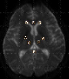
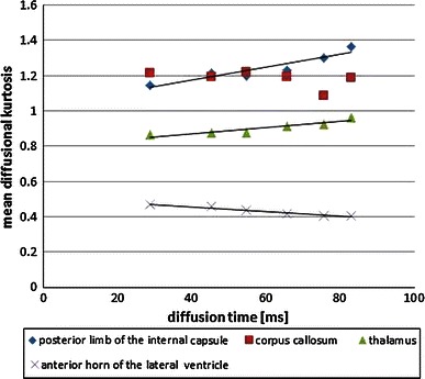
Diffusional kurtosis imaging (DKI) is a new technique based on non-Gaussian water diffusion analysis. However, the original DKI protocol (six b values and 30 motion-probing gradient (MPG) directions) requires more than 10 min of scanning time, which is too long for daily clinical use. We aimed to find suitable b value, MPG direction, and diffusion time settings for faster DKI. Four normal healthy subjects participated in the study. All DKI data sets were acquired on a clinical 3T-MRI scanner (Philips Medical Systems) with use of three protocols of 0-7500 s/mm(2) b values, 6-32 MPG directions, and 23-80 ms diffusion time. There was a remarkable difference in the standard deviation (SD) of the mean DK values in the number of MPG directions. The mean DK values were significantly higher in the posterior limb of the internal capsule (p = 0.003, r = 0.924) and thalamus (p = 0.005, r = 0.903), whereas the mean DK values of the cerebrospinal fluid (CSF) (p = 0.001, r = -0.976) were significantly lower when we used a longer diffusion time. Our results indicate that the SD of the mean DK values was higher in 15 MPG directions than in 20 MPG directions and more. Because the mean DK values of the CSF were significantly lower when we used longer diffusion times, we expect longer diffusion times to be useful for DKI. We propose the following imaging parameters for clinical use: 0, 1000, and 2000 s/mm(2) b values; 20 MPG directions; Δ/δ 45.3/13.3 ms.

摘要

扩散峰度成像(DKI)是一种基于非高斯水扩散分析的新技术。然而,原始的DKI方案(六个b值和30个运动探测梯度(MPG)方向)需要超过10分钟的扫描时间,这对于日常临床使用来说太长了。我们旨在找到合适的b值、MPG方向和扩散时间设置,以实现更快的DKI。四名正常健康受试者参与了该研究。所有DKI数据集均在临床3T-MRI扫描仪(飞利浦医疗系统公司)上采集,使用了三种协议,b值为0 - 7500 s/mm²,MPG方向为6 - 32个,扩散时间为23 - 80毫秒。MPG方向数量不同时,平均DK值的标准差(SD)存在显著差异。当使用较长扩散时间时,内囊后肢(p = 0.003,r = 0.924)和丘脑(p = 0.005,r = 0.903)的平均DK值显著更高,而脑脊液(CSF)的平均DK值(p = 0.001,r = -0.976)则显著更低。我们的结果表明,15个MPG方向时平均DK值的SD高于20个及更多MPG方向时。由于使用较长扩散时间时CSF的平均DK值显著更低,我们预计较长的扩散时间对DKI有用。我们提出以下临床使用的成像参数:b值为0、1000和2000 s/mm²;20个MPG方向;Δ/δ为45.3/13.3毫秒。

https://cdn.ncbi.nlm.nih.gov/pmc/blobs/c378/3709076/6af16f2806b7/12194_2013_206_Fig3_HTML.jpg
https://cdn.ncbi.nlm.nih.gov/pmc/blobs/c378/3709076/2f8488409b66/12194_2013_206_Fig1_HTML.jpg
https://cdn.ncbi.nlm.nih.gov/pmc/blobs/c378/3709076/70139e107f7a/12194_2013_206_Fig2_HTML.jpg
https://cdn.ncbi.nlm.nih.gov/pmc/blobs/c378/3709076/6af16f2806b7/12194_2013_206_Fig3_HTML.jpg
https://cdn.ncbi.nlm.nih.gov/pmc/blobs/c378/3709076/2f8488409b66/12194_2013_206_Fig1_HTML.jpg
https://cdn.ncbi.nlm.nih.gov/pmc/blobs/c378/3709076/70139e107f7a/12194_2013_206_Fig2_HTML.jpg
https://cdn.ncbi.nlm.nih.gov/pmc/blobs/c378/3709076/6af16f2806b7/12194_2013_206_Fig3_HTML.jpg

相似文献

1
Effects of diffusional kurtosis imaging parameters on diffusion quantification.扩散峰度成像参数对扩散量化的影响。
Radiol Phys Technol. 2013 Jul;6(2):343-8. doi: 10.1007/s12194-013-0206-5. Epub 2013 Mar 28.
2
MRI quantification of non-Gaussian water diffusion by kurtosis analysis.磁共振成像(MRI)通过峰度分析对非高斯水扩散的定量研究。
NMR Biomed. 2010 Aug;23(7):698-710. doi: 10.1002/nbm.1518.
3
Effect of cerebral spinal fluid suppression for diffusional kurtosis imaging.弥散峰度成像中脑脊液抑制的效果。
J Magn Reson Imaging. 2013 Feb;37(2):365-71. doi: 10.1002/jmri.23840. Epub 2012 Oct 3.
4
Differences in Gaussian diffusion tensor imaging and non-Gaussian diffusion kurtosis imaging model-based estimates of diffusion tensor invariants in the human brain.基于高斯扩散张量成像和非高斯扩散峰度成像模型的人脑扩散张量不变量估计差异。
Med Phys. 2016 May;43(5):2464. doi: 10.1118/1.4946819.
5
Diffusional kurtosis imaging in head and neck cancer: On the use of trace-weighted images to estimate indices of non-Gaussian water diffusion.头颈部癌症的扩散峰度成像:利用示踪加权图像估计非高斯水分子扩散指数。
Med Phys. 2018 Dec;45(12):5411-5419. doi: 10.1002/mp.13238. Epub 2018 Nov 8.
6
Fast and robust estimation of diffusional kurtosis imaging (DKI) parameters by general closed-form expressions and their extensions.通过通用闭式表达式及其扩展对扩散峰度成像(DKI)参数进行快速且稳健的估计。
Magn Reson Med Sci. 2014;13(2):97-115. doi: 10.2463/mrms.2013-0084. Epub 2014 Apr 28.
7
Diffusion-tensor-based method for robust and practical estimation of axial and radial diffusional kurtosis.基于扩散张量的轴向和径向扩散峰度稳健实用估计方法。
Eur Radiol. 2016 Aug;26(8):2559-66. doi: 10.1007/s00330-015-4038-z. Epub 2015 Oct 7.
8
Evaluating High Spatial Resolution Diffusion Kurtosis Imaging at 3T: Reproducibility and Quality of Fit.评估 3T 下高空间分辨率扩散峰度成像的可重复性和拟合质量。
J Magn Reson Imaging. 2021 Apr;53(4):1175-1187. doi: 10.1002/jmri.27408. Epub 2020 Oct 24.
9
Diffusion kurtosis imaging does not improve differentiation performance of breast lesions in a short clinical protocol.扩散峰度成像在短期临床方案中不能提高乳腺病变的鉴别性能。
Magn Reson Imaging. 2019 Nov;63:205-216. doi: 10.1016/j.mri.2019.08.007. Epub 2019 Aug 16.
10
In vivo microscopic diffusional kurtosis imaging with symmetrized double diffusion encoding EPI.对称双扩散编码 EPI 的活体微观扩散峰度成像。
Magn Reson Med. 2019 Jan;81(1):533-541. doi: 10.1002/mrm.27419. Epub 2018 Sep 9.

引用本文的文献

1
Exploring mean kurtosis in MR diffusion kurtosis imaging for early detection of lumbar spine degeneration: a systematic review.探索磁共振扩散峰度成像中的平均峰度用于早期检测腰椎退变:一项系统综述
F1000Res. 2025 Apr 15;14:440. doi: 10.12688/f1000research.163638.1. eCollection 2025.
2
Robust frequency-dependent diffusional kurtosis computation using an efficient direction scheme, axisymmetric modelling, and spatial regularization.使用高效方向方案、轴对称建模和空间正则化进行稳健的频率依赖性扩散峰度计算。
Imaging Neurosci (Camb). 2024 Jan 5;2. doi: 10.1162/imag_a_00055. eCollection 2024.
3
An Assessment of the Diagnostic Accuracy of Magnetic Resonance Diffusion Kurtosis Imaging in Diagnosing Degenerative Lumbar Spine Disease: A Scoping Review.

本文引用的文献

1
Visualizing non-Gaussian diffusion: clinical application of q-space imaging and diffusional kurtosis imaging of the brain and spine.可视化非高斯扩散:脑和脊柱 q 空间成像及扩散峰度成像的临床应用。
Magn Reson Med Sci. 2012;11(4):221-33. doi: 10.2463/mrms.11.221.
2
Regional values of diffusional kurtosis estimates in the healthy brain.健康脑区扩散峰度值的区域差异。
J Magn Reson Imaging. 2013 Mar;37(3):610-8. doi: 10.1002/jmri.23857. Epub 2012 Oct 10.
3
Effect of cerebral spinal fluid suppression for diffusional kurtosis imaging.
磁共振扩散峰度成像诊断退行性腰椎疾病的诊断准确性评估:一项范围综述
Med J Islam Repub Iran. 2025 Apr 21;39:57. doi: 10.47176/mjiri.39.57. eCollection 2025.
4
A narrative review of diffusion kurtosis MRI parameters in diagnosing degenerative spine diseases in animal models.扩散峰度磁共振成像参数在动物模型中诊断退行性脊柱疾病的叙述性综述。
Open Vet J. 2024 Dec;14(12):3181-3188. doi: 10.5455/OVJ.2024.v14.i12.3. Epub 2024 Dec 31.
5
Impact of truncating diffusion MRI scans on diffusional kurtosis imaging.截断弥散磁共振成像扫描对弥散峰度成像的影响。
MAGMA. 2024 Oct;37(5):859-872. doi: 10.1007/s10334-024-01153-y. Epub 2024 Feb 23.
6
Optimization of scan parameters to reduce acquisition time for RESOLVE-based diffusion kurtosis imaging (DKI) in nasopharyngeal carcinoma (NPC).优化扫描参数以减少基于 RESOLVE 的鼻咽癌扩散峰度成像(DKI)的采集时间。
Br J Radiol. 2022 Aug 1;95(1136):20210641. doi: 10.1259/bjr.20210641. Epub 2022 Jun 22.
7
Development of voxel-based optimization diffusion kurtosis imaging (DKI) and comparison with conventional DKI.基于体素的优化扩散峰度成像(DKI)的发展及其与传统DKI的比较。
Radiol Phys Technol. 2019 Sep;12(3):290-298. doi: 10.1007/s12194-019-00523-9. Epub 2019 Jul 5.
8
Multiparametric Functional Magnetic Resonance Imaging for Evaluating Renal Allograft Injury.多参数功能磁共振成像评估肾移植损伤。
Korean J Radiol. 2019 Jun;20(6):894-908. doi: 10.3348/kjr.2018.0540.
9
In vivo Diffusion Tensor Imaging, Diffusion Kurtosis Imaging, and Tractography of a Sciatic Nerve Injury Model in Rat at 9.4T.在 9.4T 磁共振成像系统下对大鼠坐骨神经损伤模型的体内扩散张量成像、扩散峰度成像和束流追踪研究
Sci Rep. 2018 Aug 27;8(1):12911. doi: 10.1038/s41598-018-30961-1.
10
Diffusion Kurtosis Imaging and Pathology in Spinal Cord Ischemia/Reperfusion Injury in Rabbits: A Case-Control Study.弥散峰度成像与兔脊髓缺血再灌注损伤的病理对照研究。
Med Sci Monit. 2017 Aug 18;23:3996-4003. doi: 10.12659/msm.902986.
弥散峰度成像中脑脊液抑制的效果。
J Magn Reson Imaging. 2013 Feb;37(2):365-71. doi: 10.1002/jmri.23840. Epub 2012 Oct 3.
4
New diffusion metrics for spondylotic myelopathy at an early clinical stage.早期临床阶段的颈椎病新扩散指标。
Eur Radiol. 2012 Aug;22(8):1797-802. doi: 10.1007/s00330-012-2410-9. Epub 2012 Mar 13.
5
Parkinson disease: diagnostic utility of diffusion kurtosis imaging.帕金森病:扩散峰度成像的诊断效用。
Radiology. 2011 Oct;261(1):210-7. doi: 10.1148/radiol.11102277. Epub 2011 Jul 19.
6
MR diffusion imaging in ischemic stroke.磁共振弥散成像在缺血性脑卒中的应用。
Neuroimaging Clin N Am. 2011 May;21(2):345-77, xi. doi: 10.1016/j.nic.2011.03.001.
7
Preliminary evidence of altered gray and white matter microstructural development in the frontal lobe of adolescents with attention-deficit hyperactivity disorder: a diffusional kurtosis imaging study.注意缺陷多动障碍青少年额叶灰质和白质微观结构发育改变的初步证据:扩散峰度成像研究。
J Magn Reson Imaging. 2011 Jan;33(1):17-23. doi: 10.1002/jmri.22397.
8
[Influence of imaging parameters on the measurement of apparent diffusion coefficient].[成像参数对表观扩散系数测量的影响]
Nihon Hoshasen Gijutsu Gakkai Zasshi. 2010 Sep 20;66(9):1178-85. doi: 10.6009/jjrt.66.1178.
9
MRI quantification of non-Gaussian water diffusion by kurtosis analysis.磁共振成像(MRI)通过峰度分析对非高斯水扩散的定量研究。
NMR Biomed. 2010 Aug;23(7):698-710. doi: 10.1002/nbm.1518.
10
Cerebral gliomas: diffusional kurtosis imaging analysis of microstructural differences.脑胶质瘤:扩散峰度成像分析的微观结构差异。
Radiology. 2010 Mar;254(3):876-81. doi: 10.1148/radiol.09090819. Epub 2010 Jan 20.