Division of Bacterial, Parasitic, and Allergenic Products, Center for Biologics Evaluation and Research, Food and Drug Administration, Bethesda, MD, USA.
PLoS One. 2013;8(3):e59232. doi: 10.1371/journal.pone.0059232. Epub 2013 Mar 12.
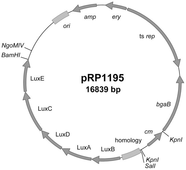
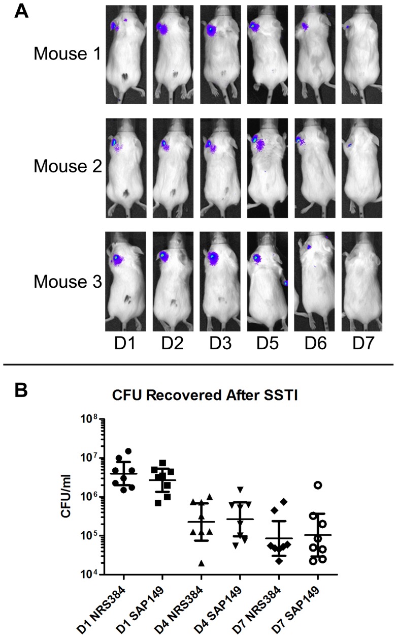
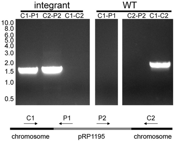
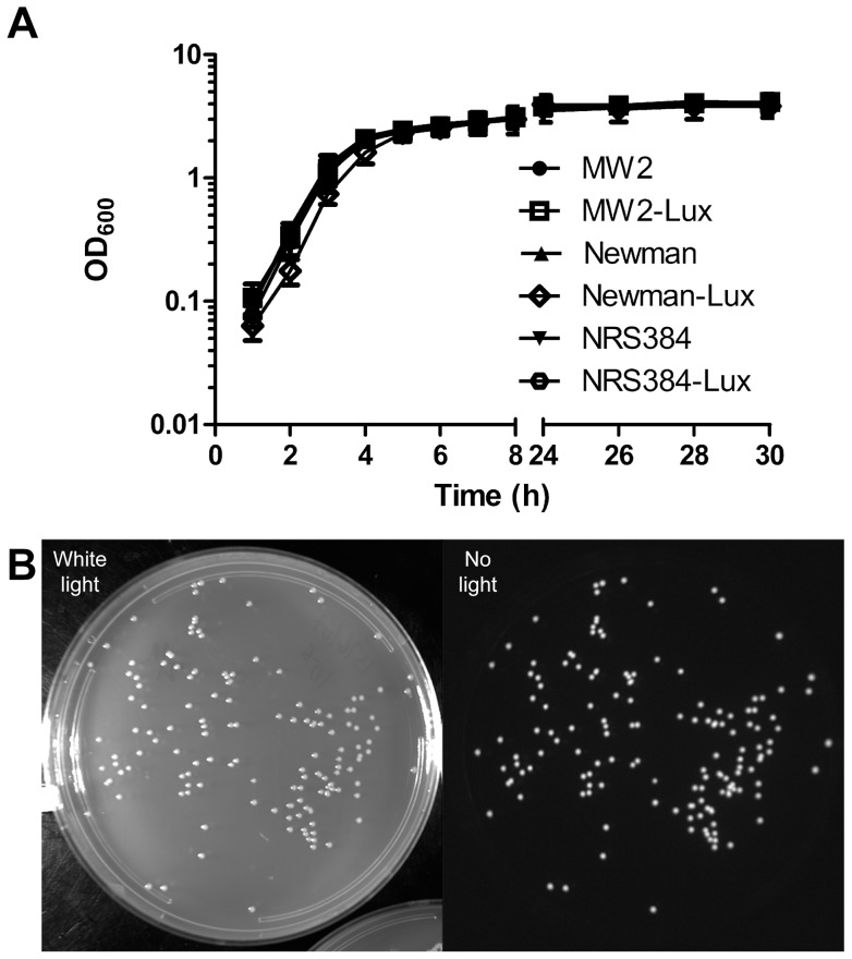
In vivo bioluminescent imaging permits the visualization of bacteria in live animals, allowing researchers to monitor, both temporally and spatially, the progression of infection in each animal. We sought to engineer stably luminescent clinical strains of Staphylococcus aureus, with the goal of using such strains in mouse models. The gram-positive shuttle vector pMAD was used as the backbone for an integration plasmid. A chloramphenicol resistance gene, a modified lux operon from Photorhabdus luminescens, and approximately 650 bp of homology to the chromosome of the USA300 S. aureus strain NRS384 were added, generating plasmid pRP1195. Electroporation into strain RN4220 followed by temperature shift led to integration of pRP1195 into the chromosome. The integrated plasmid was transferred to clinical strains by phage transduction. Luminescent strains displayed no in vitro growth defects. Moreover, luminescence was stable in vitro after three rounds of subculture over 48 hours of growth in the absence of antibiotics. Mice were infected with a luminescent strain of NRS384 in skin and intravenous models. In a mouse skin model, luminescent bacteria were present in lesions that formed and cleared over the course of several days, and in an intravenous model, bacteria inoculated in the mouse tail vein were observed spreading to multiple tissues. No statistically significant difference in virulence was observed between NRS384 and the luminescent strain in either infection model. These preliminary data suggest that this luminescent USA300 strain is suitable for use in mouse models. Similar strains were engineered using other sequenced clinical strains. Because these strains are stably luminescent, they should prove useful in animal models of infection.
在体生物发光成像是一种可以在活体动物中可视化细菌的方法,它使研究人员能够在时间和空间上监测每只动物感染的进展情况。我们试图构建稳定发光的临床金黄色葡萄球菌菌株,目的是在小鼠模型中使用这些菌株。革兰氏阳性穿梭载体 pMAD 被用作整合质粒的骨架。加入了氯霉素抗性基因、来自 Photorhabdus luminescens 的改良 lux 操纵子和大约 650bp 与 USA300 金黄色葡萄球菌 NRS384 染色体的同源性,从而产生质粒 pRP1195。将电穿孔转入 RN4220 菌株,然后进行温度转换,导致 pRP1195 整合到染色体中。通过噬菌体转导将整合质粒转移到临床菌株。发光菌株在体外没有生长缺陷。此外,在没有抗生素的情况下,经过 48 小时的生长,连续传代 3 次后,体外发光仍然稳定。用 NRS384 的发光菌株感染皮肤和静脉感染模型的小鼠。在小鼠皮肤模型中,发光细菌存在于形成和清除的病变中,持续数天;在静脉感染模型中,观察到接种于小鼠尾静脉的细菌扩散到多个组织。在两种感染模型中,NRS384 和发光菌株的毒力均无统计学差异。这些初步数据表明,这种发光的 USA300 菌株适合在小鼠模型中使用。使用其他测序的临床菌株构建了类似的菌株。由于这些菌株是稳定发光的,它们应该在感染动物模型中很有用。