Vanderbilt University Institute of Imaging Science, Nashville, Tennessee, USA.
PLoS One. 2013 Apr 26;8(4):e62708. doi: 10.1371/journal.pone.0062708. Print 2013.
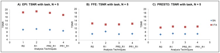
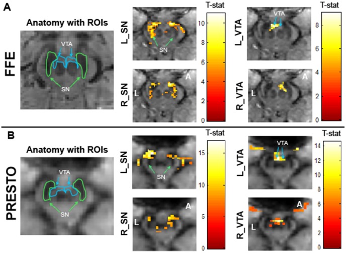
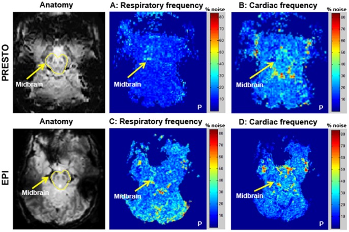
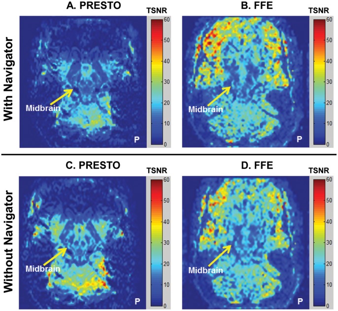
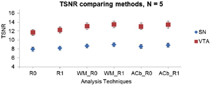
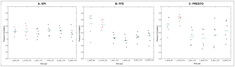
Functional Magnetic Resonance Imaging (fMRI) in the midbrain at 7 Tesla suffers from unexpectedly low temporal signal to noise ratio (TSNR) compared to other brain regions. Various methodologies were used in this study to quantitatively identify causes of the noise and signal differences in midbrain fMRI data. The influence of physiological noise sources was examined using RETROICOR, phase regression analysis, and power spectral analyses of contributions in the respiratory and cardiac frequency ranges. The impact of between-shot phase shifts in 3-D multi-shot sequences was tested using a one-dimensional (1-D) phase navigator approach. Additionally, the effects of shared noise influences between regions that were temporally, but not functionally, correlated with the midbrain (adjacent white matter and anterior cerebellum) were investigated via analyses with regressors of 'no interest'. These attempts to reduce noise did not improve the overall TSNR in the midbrain. In addition, the steady state signal and noise were measured in the midbrain and the visual cortex for resting state data. We observed comparable steady state signals from both the midbrain and the cortex. However, the noise was 2-3 times higher in the midbrain relative to the cortex, confirming that the low TSNR in the midbrain was not due to low signal but rather a result of large signal variance. These temporal variations did not behave as known physiological or other noise sources, and were not mitigated by conventional strategies. Upon further investigation, resting state functional connectivity analysis in the midbrain showed strong intrinsic fluctuations between homologous midbrain regions. These data suggest that the low TSNR in the midbrain may originate from larger signal fluctuations arising from functional connectivity compared to cortex, rather than simply reflecting physiological noise.
7 特斯拉下中脑功能磁共振成像(fMRI)的时间信号噪声比(TSNR)出乎意料地低于其他脑区。本研究采用多种方法对中脑 fMRI 数据中噪声和信号差异的原因进行定量分析。使用 RETROICOR、相位回归分析以及呼吸和心脏频率范围内的贡献的功率谱分析来检查生理噪声源的影响。通过一维(1-D)相位导航方法测试 3-D 多shot 序列中 shot 间相位偏移的影响。此外,通过分析与中脑(相邻白质和前小脑)在时间上但无功能相关的“无兴趣”回归器,研究了在时间上但与中脑不相关的区域之间共享噪声影响的影响。这些降低噪声的尝试并没有提高中脑的整体 TSNR。此外,还测量了静息状态数据中中脑和视觉皮层的稳态信号和噪声。我们观察到从中脑和皮层都可以获得可比的稳态信号。然而,中脑的噪声比皮层高 2-3 倍,这证实了中脑的低 TSNR 不是由于信号低,而是由于信号方差大所致。这些时间变化的行为与已知的生理或其他噪声源不同,并且常规策略无法减轻。进一步研究表明,中脑的静息状态功能连接分析显示出同源中脑区域之间的强烈固有波动。这些数据表明,与皮层相比,中脑的低 TSNR 可能源于功能连接引起的更大信号波动,而不仅仅反映生理噪声。