Institute of Medical Psychology, Medical Faculty, Otto-von-Guericke University of Magdeburg, Magdeburg, Germany.
PLoS One. 2013 May 8;8(5):e63700. doi: 10.1371/journal.pone.0063700. Print 2013.
Damage along the visual pathway results in a visual field defect (scotoma), which retinotopically corresponds to the damaged neural tissue. Other parts of the visual field, processed by the uninjured tissue, are considered to be intact. However, perceptual deficits have been observed in the "intact" visual field, but these functional impairments are poorly understood. We now studied temporal processing deficits in the intact visual field of patients with either pre- or post-chiasmatic lesions to better understand the functional consequences of partial blindness.
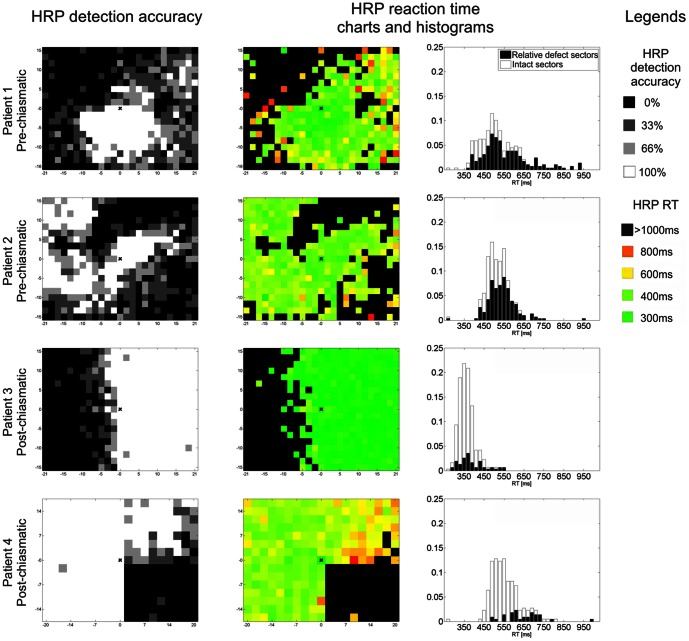
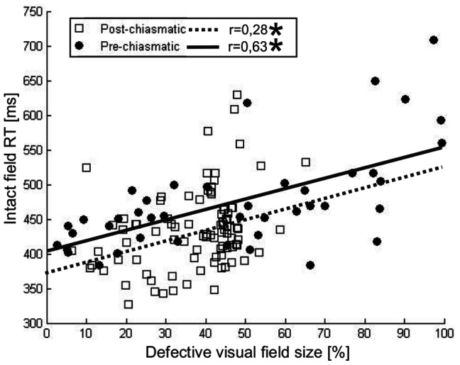
Patients with pre- (n = 53) or post-chiasmatic lesions (n = 98) were tested with high resolution perimetry--a method used to map visual fields with supra-threshold light stimuli. Reaction time of detections in the intact visual field was then analyzed as an indicator of processing speed and correlated with features of the visual field defect.
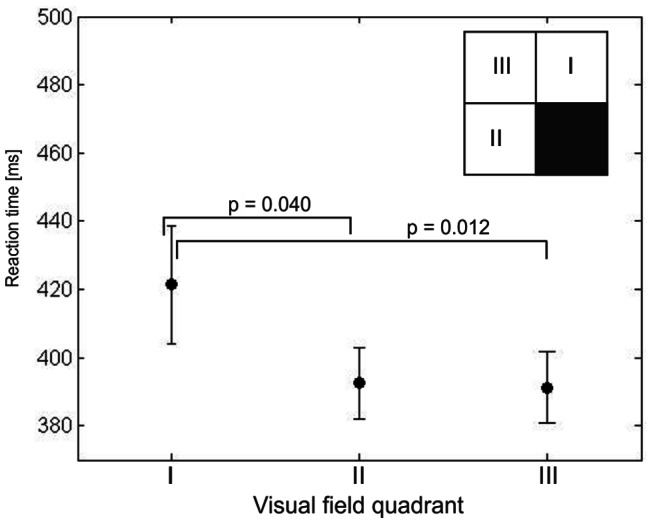
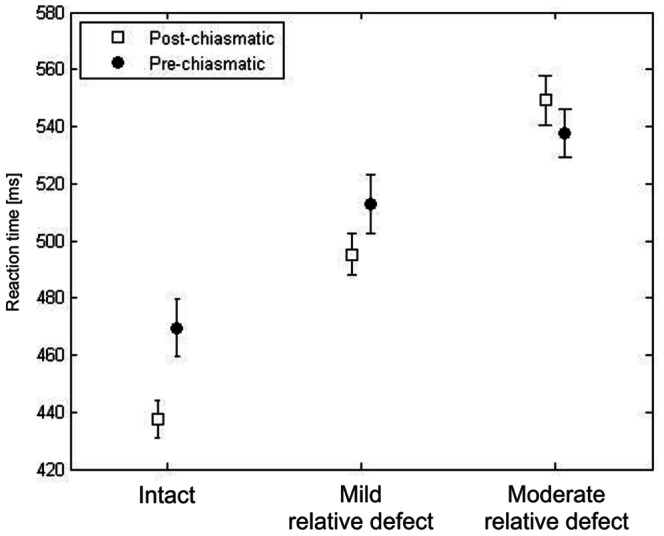
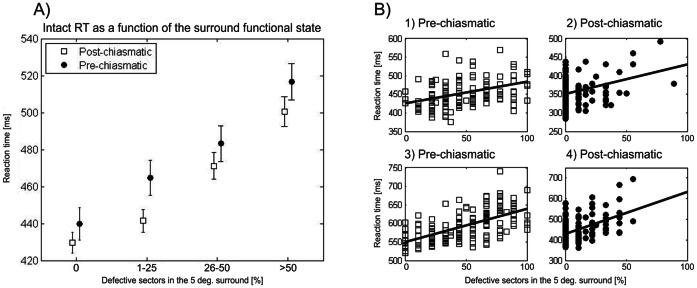
Patients from both groups exhibited processing speed deficits in their presumably "intact" field as indicated by comparison to a normative sample. Further, in both groups processing speed was found to be a function of two factors. Firstly, a spatially restricted (retinotopic) influence of the scotoma was seen in longer reaction times when stimuli were presented in intact field sectors close to the defect. Secondly, patients with larger scotomata had on average longer reaction times in their intact field indicating a more general (non-retinotopic) influence of the scotoma.
Processing speed deficits in the "intact" visual field of patients with visual system damage demonstrate that visual system lesions have more widespread consequences on perception than previously thought. Because dysfunctions of the seeing field are expected to contribute to subjective vision, including visual tests of the presumed "intact" field may help to better understand vision loss and to improve methods of vision restoration and rehabilitation.
视觉通路损伤会导致视野缺损(暗点),其与损伤的神经组织在视网膜上相对应。其他由未受损组织处理的视野部分被认为是完整的。然而,在“完整”视野中已经观察到知觉缺陷,但这些功能障碍的理解较差。我们现在研究了视交叉前或视交叉后病变患者的完整视野中的时间处理缺陷,以更好地理解部分失明的功能后果。
使用高分辨率视野计测试视交叉前病变(n=53)或视交叉后病变患者(n=98)-一种用于用超阈值光刺激绘制视野的方法。然后分析完整视野中的检测反应时间作为处理速度的指标,并与视野缺陷的特征相关联。
两组患者在其“完整”视野中均表现出处理速度缺陷,与正常样本相比,这表明了这一点。此外,在两组中,处理速度被发现是两个因素的函数。首先,在刺激呈现于接近缺损的完整视野区域时,视盲的空间限制(视网膜)影响导致反应时间延长。其次,视盲较大的患者在其完整视野中的反应时间平均更长,这表明视盲具有更普遍的(非视网膜)影响。
视觉系统损伤患者的“完整”视野中的处理速度缺陷表明,视觉系统损伤对感知的影响比以前认为的更为广泛。由于视场功能障碍预计会对视觉产生主观影响,包括对假定“完整”视野的视觉测试,可能有助于更好地理解视力丧失,并改善视力恢复和康复方法。