• 文献检索
  • 文档翻译
  • 深度研究
  • 学术资讯
  • Suppr Zotero 插件Zotero 插件
  • 邀请有礼
  • 套餐&价格
  • 历史记录
应用&插件
Suppr Zotero 插件Zotero 插件浏览器插件Mac 客户端Windows 客户端微信小程序
定价
高级版会员购买积分包购买API积分包
服务
文献检索文档翻译深度研究API 文档MCP 服务
关于我们
关于 Suppr公司介绍联系我们用户协议隐私条款
关注我们

Suppr 超能文献

核心技术专利:CN118964589B侵权必究
粤ICP备2023148730 号-1Suppr @ 2026

文献检索

告别复杂PubMed语法,用中文像聊天一样搜索,搜遍4000万医学文献。AI智能推荐,让科研检索更轻松。

立即免费搜索

文件翻译

保留排版,准确专业,支持PDF/Word/PPT等文件格式,支持 12+语言互译。

免费翻译文档

深度研究

AI帮你快速写综述,25分钟生成高质量综述,智能提取关键信息,辅助科研写作。

立即免费体验

利用高角度分辨率扩散成像数据对皮质区域进行区分。

Using high angular resolution diffusion imaging data to discriminate cortical regions.

机构信息

Wellcome Trust Centre for Neuroimaging, UCL Institute of Neurology, University College London, London, United Kingdom.

出版信息

PLoS One. 2013 May 17;8(5):e63842. doi: 10.1371/journal.pone.0063842. Print 2013.

DOI:10.1371/journal.pone.0063842
PMID:23691102
原文链接:https://pmc.ncbi.nlm.nih.gov/articles/PMC3656939/
Abstract

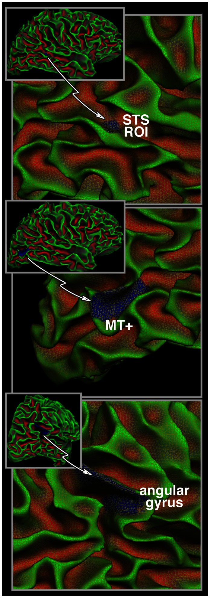
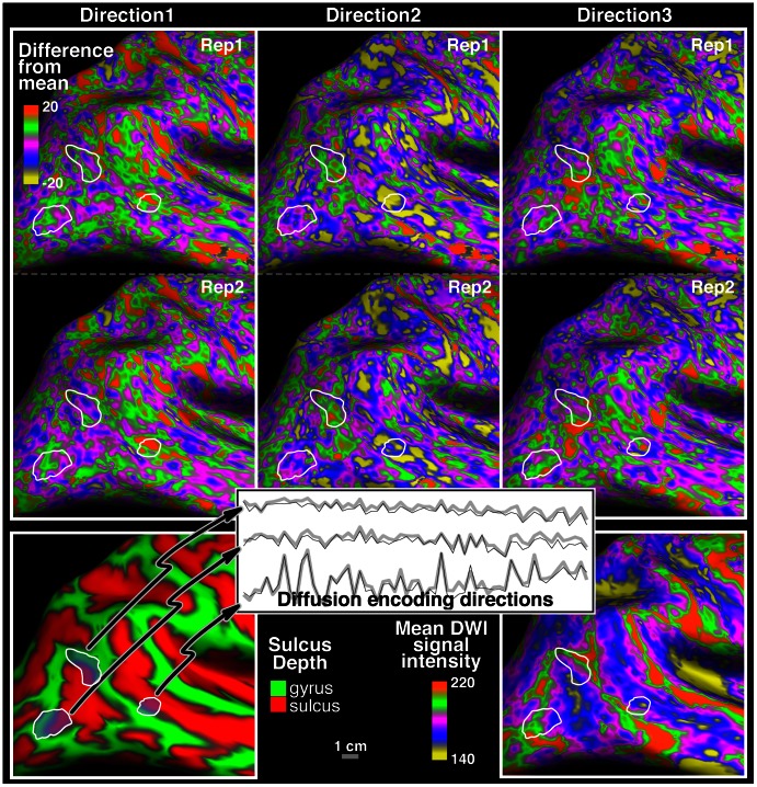
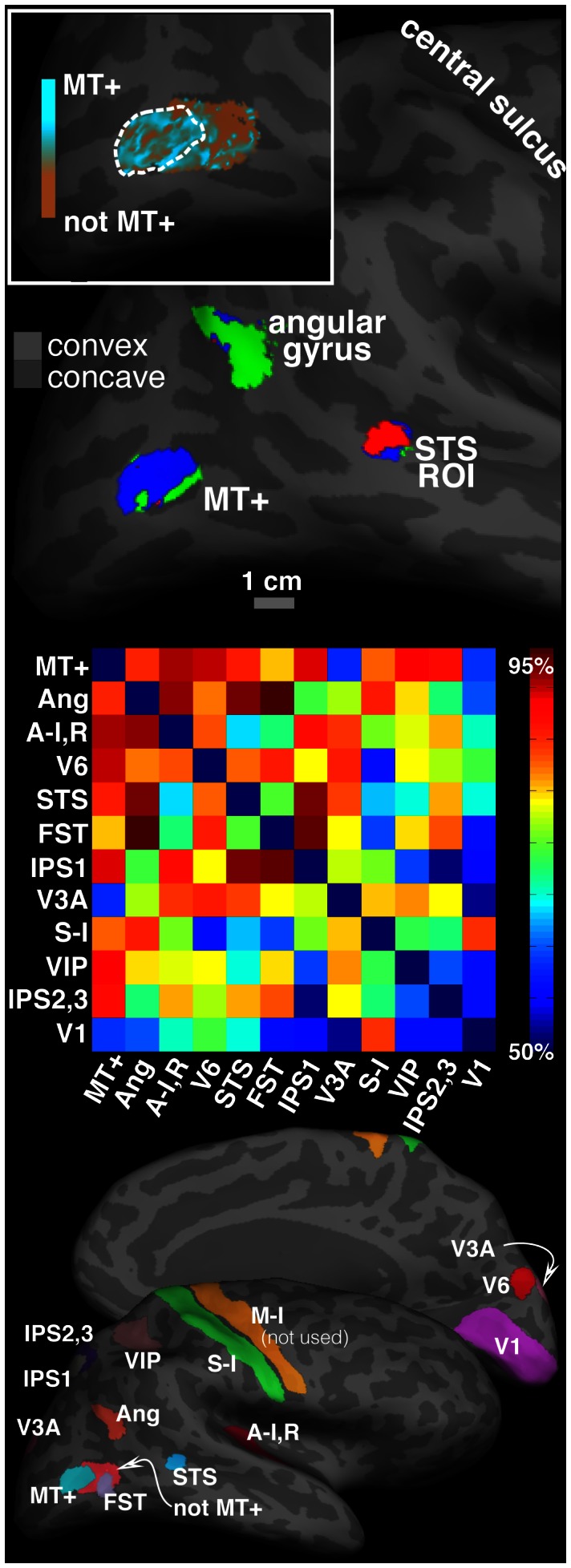
Brodmann's 100-year-old summary map has been widely used for cortical localization in neuroscience. There is a pressing need to update this map using non-invasive, high-resolution and reproducible data, in a way that captures individual variability. We demonstrate here that standard HARDI data has sufficiently diverse directional variation among grey matter regions to inform parcellation into distinct functional regions, and that this variation is reproducible across scans. This characterization of the signal variation as non-random and reproducible is the critical condition for successful cortical parcellation using HARDI data. This paper is a first step towards an individual cortex-wide map of grey matter microstructure, The gray/white matter and pial boundaries were identified on the high-resolution structural MRI images. Two HARDI data sets were collected from each individual and aligned with the corresponding structural image. At each vertex point on the surface tessellation, the diffusion-weighted signal was extracted from each image in the HARDI data set at a point, half way between gray/white matter and pial boundaries. We then derived several features of the HARDI profile with respect to the local cortical normal direction, as well as several fully orientationally invariant features. These features were taken as a fingerprint of the underlying grey matter tissue, and used to distinguish separate cortical areas. A support-vector machine classifier, trained on three distinct areas in repeat 1 achieved 80-82% correct classification of the same three areas in the unseen data from repeat 2 in three volunteers. Though gray matter anisotropy has been mostly overlooked hitherto, this approach may eventually form the foundation of a new cortical parcellation method in living humans. Our approach allows for further studies on the consistency of HARDI based parcellation across subjects and comparison with independent microstructural measures such as ex-vivo histology.

摘要

布罗德曼 100 年前的综述图在神经科学中被广泛用于皮质定位。现在迫切需要使用非侵入性、高分辨率和可重复的数据来更新该图谱,以捕捉个体差异。我们在此证明,标准 HARDI 数据在灰质区域之间具有足够多样化的方向变化,可以将其划分为不同的功能区域,并且这种变化在扫描之间是可重复的。这种将信号变化描述为非随机和可重复的特征是使用 HARDI 数据进行成功皮质分割的关键条件。本文是朝着个体皮质全灰质微观结构图谱迈出的第一步。高分辨率结构 MRI 图像确定了灰质/白质和软脑膜边界。从每个个体收集了两个 HARDI 数据集,并与相应的结构图像对齐。在表面剖分的每个顶点处,从 HARDI 数据集中的每个图像在灰质/白质和软脑膜边界之间的中点提取扩散加权信号。然后,我们根据局部皮质法向方向推导出 HARDI 轮廓的几个特征,以及几个完全各向同性不变的特征。这些特征被视为潜在灰质组织的指纹,并用于区分不同的皮质区域。在三个志愿者的重复 1 中的三个不同区域上训练的支持向量机分类器,在重复 2 中的未见过数据中对相同的三个区域实现了 80-82%的正确分类。尽管迄今为止灰质各向异性大多被忽视,但这种方法最终可能成为活体人类新皮质分割方法的基础。我们的方法允许进一步研究 HARDI 分割在不同受试者之间的一致性,并与独立的微观结构测量(如离体组织学)进行比较。

https://cdn.ncbi.nlm.nih.gov/pmc/blobs/e0e4/3656939/8641b04a3c61/pone.0063842.g004.jpg
https://cdn.ncbi.nlm.nih.gov/pmc/blobs/e0e4/3656939/371c42d3bb87/pone.0063842.g001.jpg
https://cdn.ncbi.nlm.nih.gov/pmc/blobs/e0e4/3656939/ffc38cb1bc81/pone.0063842.g002.jpg
https://cdn.ncbi.nlm.nih.gov/pmc/blobs/e0e4/3656939/99335af60167/pone.0063842.g003.jpg
https://cdn.ncbi.nlm.nih.gov/pmc/blobs/e0e4/3656939/8641b04a3c61/pone.0063842.g004.jpg

相似文献

1
Using high angular resolution diffusion imaging data to discriminate cortical regions.利用高角度分辨率扩散成像数据对皮质区域进行区分。
PLoS One. 2013 May 17;8(5):e63842. doi: 10.1371/journal.pone.0063842. Print 2013.
2
Using diffusion MRI to discriminate areas of cortical grey matter.利用弥散磁共振成像区分皮质灰质区域。
Neuroimage. 2018 Nov 15;182:456-468. doi: 10.1016/j.neuroimage.2017.12.046. Epub 2017 Dec 21.
3
Probing region-specific microstructure of human cortical areas using high angular and spatial resolution diffusion MRI.利用高角度和空间分辨率扩散 MRI 探测人类皮质区的特定区域微观结构。
Neuroimage. 2015 Jan 15;105:198-207. doi: 10.1016/j.neuroimage.2014.10.053. Epub 2014 Oct 31.
4
Group-wise consistent cortical parcellation based on connectional profiles.基于连接模式的组内一致皮质分块
Med Image Anal. 2016 Aug;32:32-45. doi: 10.1016/j.media.2016.02.009. Epub 2016 Mar 14.
5
HIgh b-value and high Resolution Integrated Diffusion (HIBRID) imaging.高b值与高分辨率综合扩散(HIBRID)成像
Neuroimage. 2017 Apr 15;150:162-176. doi: 10.1016/j.neuroimage.2017.02.002. Epub 2017 Feb 7.
6
Human brain atlas for automated region of interest selection in quantitative susceptibility mapping: application to determine iron content in deep gray matter structures.人脑图谱用于定量磁化率映射中感兴趣区的自动选择:在确定深部灰质结构铁含量中的应用。
Neuroimage. 2013 Nov 15;82:449-69. doi: 10.1016/j.neuroimage.2013.05.127. Epub 2013 Jun 12.
7
Surface based analysis of diffusion orientation for identifying architectonic domains in the in vivo human cortex.基于表面的扩散方向分析用于识别活体人脑皮质的结构域。
Neuroimage. 2013 Apr 1;69:87-100. doi: 10.1016/j.neuroimage.2012.11.065. Epub 2012 Dec 16.
8
In vivo cortical microstructure mapping using high-gradient diffusion MRI accounting for intercompartmental water exchange effects.利用考虑隔室间水交换效应的高梯度扩散磁共振成像进行体内皮质微结构映射。
Neuroimage. 2025 Jul 1;314:121258. doi: 10.1016/j.neuroimage.2025.121258. Epub 2025 May 9.
9
Fully automated classification of HARDI in vivo data using a support vector machine.使用支持向量机对体内扩散张量成像数据进行全自动分类。
Neuroimage. 2009 Jul 1;46(3):642-51. doi: 10.1016/j.neuroimage.2009.03.003. Epub 2009 Mar 12.
10
Imaging laminar structures in the gray matter with diffusion MRI.弥散 MRI 成像技术在脑灰质层状结构中的应用。
Neuroimage. 2019 Aug 15;197:677-688. doi: 10.1016/j.neuroimage.2017.12.096. Epub 2018 Jan 5.

引用本文的文献

1
Direct segmentation of cortical cytoarchitectonic domains using ultra-high-resolution whole-brain diffusion MRI.使用超高分辨率全脑扩散磁共振成像直接分割皮质细胞构筑区域
Imaging Neurosci (Camb). 2024 Dec 20;2. doi: 10.1162/imag_a_00393. eCollection 2024.
2
Direct segmentation of cortical cytoarchitectonic domains using ultra-high-resolution whole-brain diffusion MRI.使用超高分辨率全脑扩散磁共振成像直接分割皮质细胞构筑区域
bioRxiv. 2024 Oct 17:2024.10.14.618245. doi: 10.1101/2024.10.14.618245.
3
Custom integration of a magnetic-field monitoring system into a 32-channel MRI head coil.

本文引用的文献

1
Using high-resolution quantitative mapping of R1 as an index of cortical myelination.利用 R1 的高分辨率定量映射作为皮质髓鞘化的指标。
Neuroimage. 2014 Jun;93 Pt 2:176-88. doi: 10.1016/j.neuroimage.2013.06.005. Epub 2013 Jun 10.
2
Quantification of anisotropy and fiber orientation in human brain histological sections.人类脑组织切片各向异性和纤维方向的量化。
Front Integr Neurosci. 2013 Feb 1;7:3. doi: 10.3389/fnint.2013.00003. eCollection 2013.
3
The impact of post-processing on spinal cord diffusion tensor imaging.后处理对脊髓弥散张量成像的影响。
将磁场监测系统定制集成到 32 通道 MRI 头部线圈中。
Magn Reson Med. 2025 Feb;93(2):889-898. doi: 10.1002/mrm.30314. Epub 2024 Sep 29.
4
Assessing biological self-organization patterns using statistical complexity characteristics: a tool for diffusion tensor imaging analysis.利用统计复杂性特征评估生物自组织模式:一种扩散张量成像分析工具。
MAGMA. 2024 Jul 28. doi: 10.1007/s10334-024-01185-4.
5
Spatiotemporal patterns of cortical microstructural maturation in children and adolescents with diffusion MRI.基于弥散磁共振成像的儿童和青少年皮质微观结构成熟的时空模式。
Hum Brain Mapp. 2024 Jan;45(1):e26528. doi: 10.1002/hbm.26528. Epub 2023 Nov 22.
6
Multi-tensor diffusion abnormalities of gray matter in an animal model of cortical dysplasia.皮质发育异常动物模型中灰质的多张量扩散异常
Front Neurol. 2023 May 5;14:1124282. doi: 10.3389/fneur.2023.1124282. eCollection 2023.
7
COnstrained Reference frame diffusion TEnsor Correlation Spectroscopic (CORTECS) MRI: A practical framework for high-resolution diffusion tensor distribution imaging.约束参考框架扩散张量相关光谱(CORTECS)磁共振成像:一种用于高分辨率扩散张量分布成像的实用框架。
Front Neurosci. 2022 Dec 15;16:1054509. doi: 10.3389/fnins.2022.1054509. eCollection 2022.
8
Cerebral cortex layer segmentation using diffusion magnetic resonance imaging in vivo with applications to laminar connections and working memory analysis.使用活体弥散磁共振成像进行大脑皮层层分割及其在层间连接和工作记忆分析中的应用。
Hum Brain Mapp. 2022 Dec 1;43(17):5220-5234. doi: 10.1002/hbm.25998. Epub 2022 Jul 1.
9
Topological Maps and Brain Computations From Low to High.从低到高的拓扑地图与大脑计算
Front Syst Neurosci. 2022 May 27;16:787737. doi: 10.3389/fnsys.2022.787737. eCollection 2022.
10
Multiple b-values improve discrimination of cortical gray matter regions using diffusion MRI: an experimental validation with a data-driven approach.多 b 值可提高弥散磁共振成像对皮质灰质区域的辨别能力:一种基于数据驱动方法的实验验证。
MAGMA. 2021 Oct;34(5):677-687. doi: 10.1007/s10334-021-00914-3. Epub 2021 Mar 12.
Neuroimage. 2013 Apr 15;70:377-85. doi: 10.1016/j.neuroimage.2012.12.058. Epub 2013 Jan 5.
4
Surface based analysis of diffusion orientation for identifying architectonic domains in the in vivo human cortex.基于表面的扩散方向分析用于识别活体人脑皮质的结构域。
Neuroimage. 2013 Apr 1;69:87-100. doi: 10.1016/j.neuroimage.2012.11.065. Epub 2012 Dec 16.
5
Mapping the human cortical surface by combining quantitative T(1) with retinotopy.通过结合定量 T1 与视皮层投射图绘制人类皮质表面图。
Cereb Cortex. 2013 Sep;23(9):2261-8. doi: 10.1093/cercor/bhs213. Epub 2012 Jul 23.
6
NODDI: practical in vivo neurite orientation dispersion and density imaging of the human brain.NODDI:人脑神经丝取向分散和密度成像的实用体内技术。
Neuroimage. 2012 Jul 16;61(4):1000-16. doi: 10.1016/j.neuroimage.2012.03.072. Epub 2012 Mar 30.
7
Robust and fast whole brain mapping of the RF transmit field B1 at 7T.在 7T 下对 RF 发射场 B1 进行稳健快速的全脑映射。
PLoS One. 2012;7(3):e32379. doi: 10.1371/journal.pone.0032379. Epub 2012 Mar 12.
8
Mapping human cortical areas in vivo based on myelin content as revealed by T1- and T2-weighted MRI.基于 T1 加权和 T2 加权 MRI 显示的髓鞘含量对人体皮质区进行体内定位。
J Neurosci. 2011 Aug 10;31(32):11597-616. doi: 10.1523/JNEUROSCI.2180-11.2011.
9
Diffusion tensor imaging and beyond.扩散张量成像及其他。
Magn Reson Med. 2011 Jun;65(6):1532-56. doi: 10.1002/mrm.22924. Epub 2011 Apr 5.
10
Microstructural Parcellation of the Human Cerebral Cortex - From Brodmann's Post-Mortem Map to in vivo Mapping with High-Field Magnetic Resonance Imaging.人类大脑皮层的微观结构分区——从布罗德曼的尸检图谱到高场磁共振成像的活体图谱
Front Hum Neurosci. 2011 Feb 18;5:19. doi: 10.3389/fnhum.2011.00019. eCollection 2011.