Department of Biophysics, School of Life Science and Technology, University of Electronic Science and Technology of China, Chengdu, Sichuan, People's Republic of China.
Int J Nanomedicine. 2013;8:1897-906. doi: 10.2147/IJN.S44997. Epub 2013 May 13.
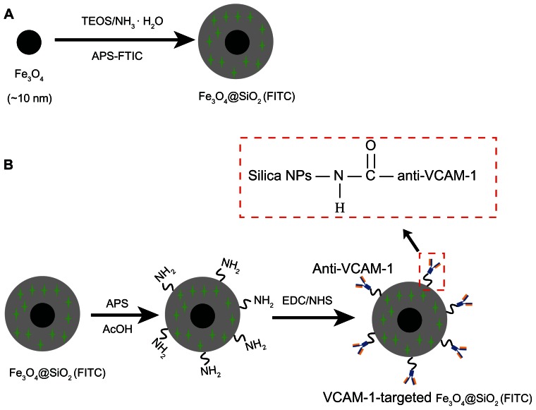
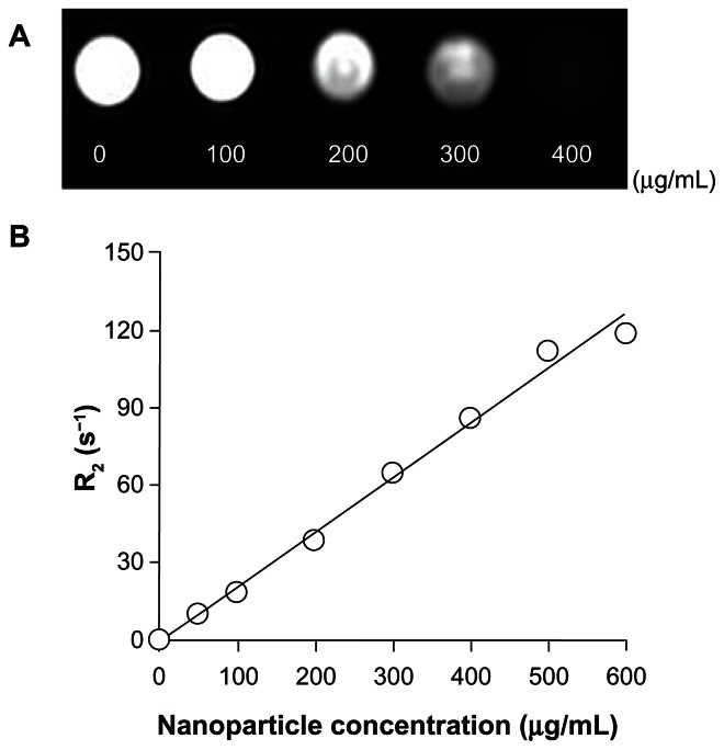
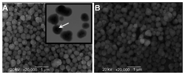
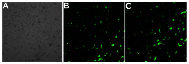
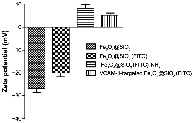
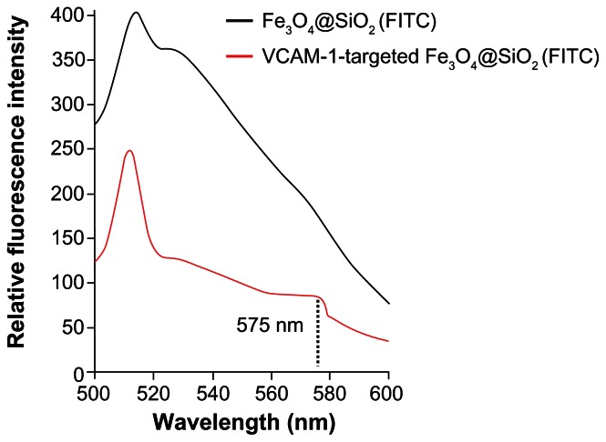
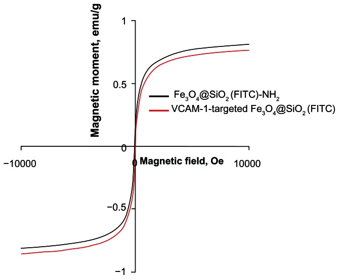
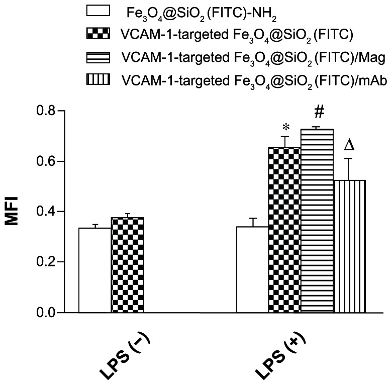
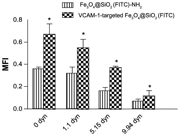
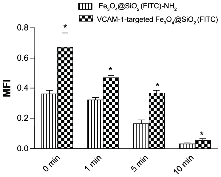
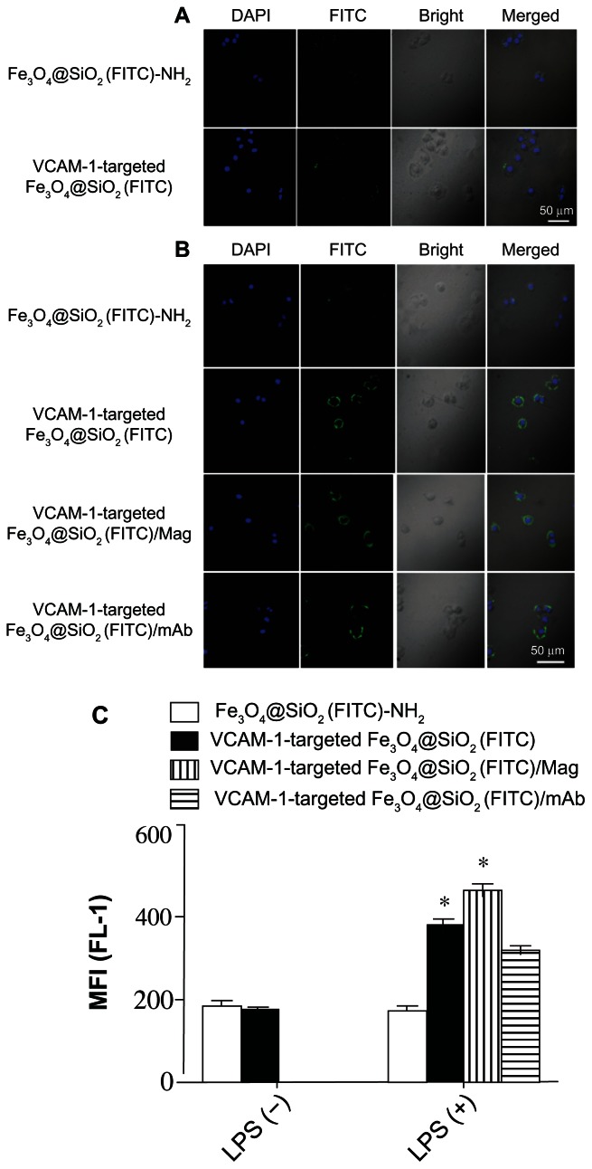
Multifunctional nanomaterials with unique magnetic and luminescent properties have broad potential in biological applications. Because of the overexpression of vascular cell adhesion molecule-1 (VCAM-1) receptors in inflammatory endothelial cells as compared with normal endothelial cells, an anti-VCAM-1 monoclonal antibody can be used as a targeting ligand. Herein we describe the development of multifunctional core-shell Fe(3)O(4)@SiO2 nanoparticles with the ability to target inflammatory endothelial cells via VCAM-1, magnetism, and fluorescence imaging, with efficient magnetic resonance imaging contrast characteristics. Superparamagnetic iron oxide and fluorescein isothiocyanate (FITC) were loaded successfully inside the nanoparticle core and the silica shell, respectively, creating VCAM-1-targeted Fe(3)O(4)@SiO2(FITC) nanoparticles that were characterized by scanning electron microscopy, transmission electron microscopy, fluorescence spectrometry, zeta potential assay, and fluorescence microscopy. The VCAM-1-targeted Fe(3)O(4)@SiO2(FITC) nanoparticles typically had a diameter of 355 ± 37 nm, showed superparamagnetic behavior at room temperature, and cumulative and targeted adhesion to an inflammatory subline of human umbilical vein endothelial cells (HUVEC-CS) activated by lipopolysaccharide. Further, our data show that adhesion of VCAM-1-targeted Fe(3)O(4)@SiO2(FITC) nanoparticles to inflammatory HUVEC-CS depended on both shear stress and duration of exposure to stress. Analysis of internalization into HUVEC-CS showed that the efficiency of delivery of VCAM-1-targeted Fe(3)O(4)@SiO2(FITC) nanoparticles was also significantly greater than that of nontargeted Fe(3)O(4)@SiO2(FITC)-NH2 nanoparticles. Magnetic resonance images showed that the superparamagnetic iron oxide cores of the VCAM-1-targeted Fe(3)O(4)@SiO2(FITC) nanoparticles could also act as a contrast agent for magnetic resonance imaging. Taken together, the cumulative adhesion and uptake potential of these VCAM-1-targeted Fe(3)O(4)@SiO2(FITC) nanoparticles targeted to inflammatory endothelial cells could be used in the transfer of therapeutic drugs/genes into these cells or for diagnosis of vascular disease at the molecular and cellular levels in the future.
具有独特磁性和发光性能的多功能纳米材料在生物应用中具有广阔的潜力。由于炎症内皮细胞中血管细胞粘附分子-1(VCAM-1)受体的过度表达,与正常内皮细胞相比,抗 VCAM-1 单克隆抗体可用作靶向配体。在此,我们描述了多功能核壳 Fe(3)O(4)@SiO2 纳米粒子的开发,该纳米粒子具有通过 VCAM-1、磁性和荧光成像靶向炎症内皮细胞的能力,具有高效的磁共振成像对比特性。超顺磁性氧化铁和异硫氰酸荧光素(FITC)分别成功地装载在纳米粒子核和二氧化硅壳内,形成靶向 VCAM-1 的 Fe(3)O(4)@SiO2(FITC)纳米粒子,通过扫描电子显微镜、透射电子显微镜、荧光光谱、Zeta 电位测定和荧光显微镜进行了表征。靶向 VCAM-1 的 Fe(3)O(4)@SiO2(FITC)纳米粒子通常直径为 355±37nm,在室温下表现出超顺磁性行为,并与脂多糖激活的炎症人脐静脉内皮细胞(HUVEC-CS)的炎症亚系累积和靶向粘附。此外,我们的数据表明,靶向 VCAM-1 的 Fe(3)O(4)@SiO2(FITC)纳米粒子与炎症 HUVEC-CS 的粘附既依赖于剪切应力,也依赖于暴露于应力的持续时间。对 HUVEC-CS 内吞作用的分析表明,靶向 VCAM-1 的 Fe(3)O(4)@SiO2(FITC)纳米粒子的递送效率也明显高于非靶向 Fe(3)O(4)@SiO2(FITC)-NH2 纳米粒子。磁共振成像显示,靶向 VCAM-1 的 Fe(3)O(4)@SiO2(FITC)纳米粒子的超顺磁性氧化铁核也可用作磁共振成像的对比剂。综上所述,这些靶向炎症内皮细胞的 VCAM-1 靶向 Fe(3)O(4)@SiO2(FITC)纳米粒子的累积粘附和摄取潜力可用于将治疗药物/基因递送到这些细胞中,或用于在分子和细胞水平上诊断血管疾病。