• 文献检索
  • 文档翻译
  • 深度研究
  • 学术资讯
  • Suppr Zotero 插件Zotero 插件
  • 邀请有礼
  • 套餐&价格
  • 历史记录
应用&插件
Suppr Zotero 插件Zotero 插件浏览器插件Mac 客户端Windows 客户端微信小程序
定价
高级版会员购买积分包购买API积分包
服务
文献检索文档翻译深度研究API 文档MCP 服务
关于我们
关于 Suppr公司介绍联系我们用户协议隐私条款
关注我们

Suppr 超能文献

核心技术专利:CN118964589B侵权必究
粤ICP备2023148730 号-1Suppr @ 2026

文献检索

告别复杂PubMed语法,用中文像聊天一样搜索,搜遍4000万医学文献。AI智能推荐,让科研检索更轻松。

立即免费搜索

文件翻译

保留排版,准确专业,支持PDF/Word/PPT等文件格式,支持 12+语言互译。

免费翻译文档

深度研究

AI帮你快速写综述,25分钟生成高质量综述,智能提取关键信息,辅助科研写作。

立即免费体验

磁共振成像对急性脑出血患者血脑屏障损伤的特征分析。

Magnetic resonance imaging profile of blood-brain barrier injury in patients with acute intracerebral hemorrhage.

机构信息

Stanford Neurocritical Care Program, Stanford Stroke Center, Stanford University Medical Center, Stanford, CA, USA.

出版信息

J Am Heart Assoc. 2013 May 24;2(3):e000161. doi: 10.1161/JAHA.113.000161.

DOI:10.1161/JAHA.113.000161
PMID:23709564
原文链接:https://pmc.ncbi.nlm.nih.gov/articles/PMC3698778/
Abstract

BACKGROUND

Spontaneous intracerebral hemorrhage (ICH) is associated with blood-brain barrier (BBB) injury, which is a poorly understood factor in ICH pathogenesis, potentially contributing to edema formation and perihematomal tissue injury. We aimed to assess and quantify BBB permeability following human spontaneous ICH using dynamic contrast-enhanced magnetic resonance imaging (DCE MRI). We also investigated whether hematoma size or location affected the amount of BBB leakage.

METHODS AND RESULTS

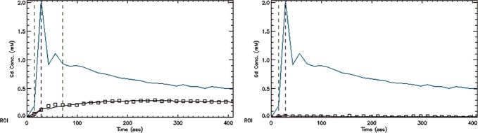
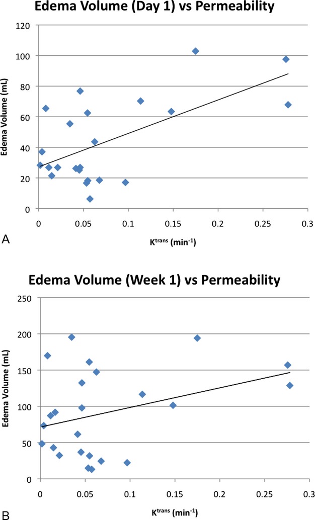
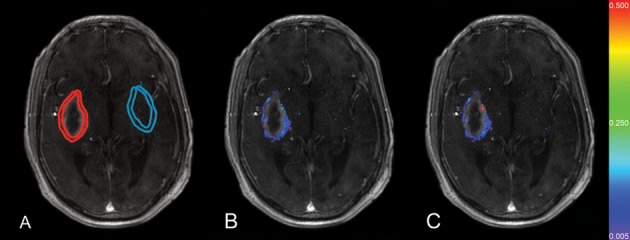
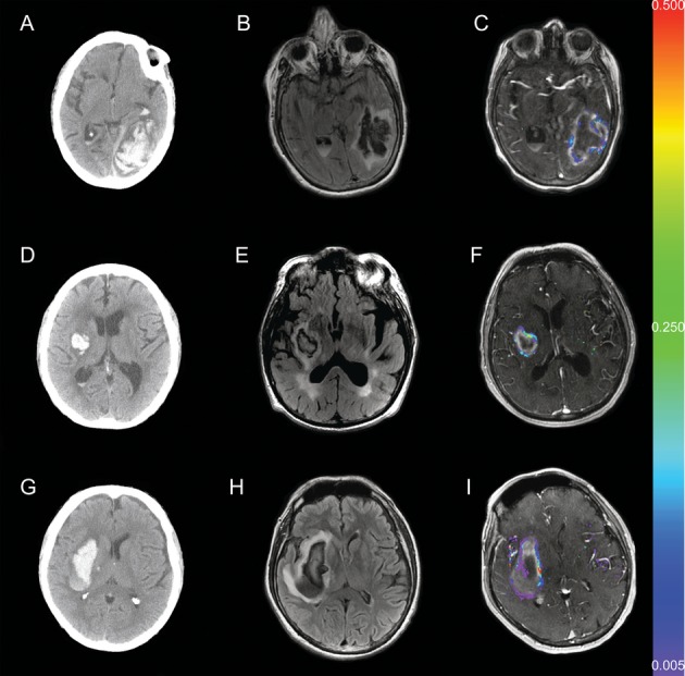
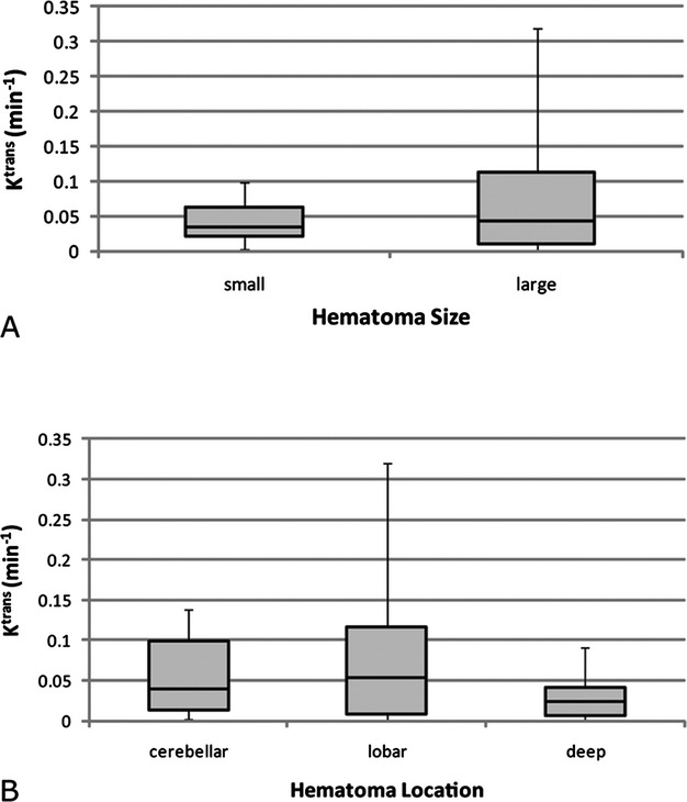
Twenty-five prospectively enrolled patients from the Diagnostic Accuracy of MRI in Spontaneous intracerebral Hemorrhage (DASH) study were examined using DCE MRI at 1 week after symptom onset. Contrast agent dynamics in the brain tissue and general tracer kinetic modeling were used to estimate the forward leakage rate (K(trans)) in regions of interest (ROI) in and surrounding the hematoma and in contralateral mirror-image locations (control ROI). In all patients BBB permeability was significantly increased in the brain tissue immediately adjacent to the hematoma, that is, the hematoma rim, compared to the contralateral mirror ROI (P<0.0001). Large hematomas (>30 mL) had higher K(trans) values than small hematomas (P<0.005). K(trans) values of lobar hemorrhages were significantly higher than the K(trans) values of deep hemorrhages (P<0.005), independent of hematoma volume. Higher K(trans) values were associated with larger edema volumes.

CONCLUSIONS

BBB leakage in the brain tissue immediately bordering the hematoma can be measured and quantified by DCE MRI in human ICH. BBB leakage at 1 week is greater in larger hematomas as well as in hematomas in lobar locations and is associated with larger edema volumes.

摘要

背景

自发性脑出血(ICH)与血脑屏障(BBB)损伤有关,后者是 ICH 发病机制中一个尚未充分了解的因素,可能导致水肿形成和血肿周围组织损伤。我们旨在使用动态对比增强磁共振成像(DCE MRI)评估和量化人自发性 ICH 后 BBB 的通透性。我们还研究了血肿大小或位置是否影响 BBB 渗漏量。

方法和结果

前瞻性纳入来自自发性脑出血的 MRI 诊断准确性研究(DASH)的 25 例患者,在发病后 1 周进行 DCE MRI 检查。使用对比剂在脑组织中的动力学和一般示踪剂动力学模型,估计 ROI 内和周围的正向漏出率(K(trans)),以及血肿对侧镜像 ROI(对照 ROI)。在所有患者中,与对侧镜像 ROI 相比,紧邻血肿的脑组织(即血肿边缘)的 BBB 通透性明显增加(P<0.0001)。大血肿(>30 mL)的 K(trans)值高于小血肿(P<0.005)。与深部出血相比,叶状出血的 K(trans)值明显更高(P<0.005),而与血肿体积无关。较高的 K(trans)值与更大的水肿体积相关。

结论

DCE MRI 可测量和量化人类 ICH 紧邻血肿的脑组织中的 BBB 渗漏。较大的血肿以及叶状位置的血肿在 1 周时的 BBB 渗漏更大,与更大的水肿体积相关。

https://cdn.ncbi.nlm.nih.gov/pmc/blobs/11d3/3698778/62e50154b3c0/jah3-2-e000161-g5.jpg
https://cdn.ncbi.nlm.nih.gov/pmc/blobs/11d3/3698778/400231322bd4/jah3-2-e000161-g1.jpg
https://cdn.ncbi.nlm.nih.gov/pmc/blobs/11d3/3698778/3d9964c8d6d5/jah3-2-e000161-g2.jpg
https://cdn.ncbi.nlm.nih.gov/pmc/blobs/11d3/3698778/f9e5fd987e2b/jah3-2-e000161-g3.jpg
https://cdn.ncbi.nlm.nih.gov/pmc/blobs/11d3/3698778/8720ab544c96/jah3-2-e000161-g4.jpg
https://cdn.ncbi.nlm.nih.gov/pmc/blobs/11d3/3698778/62e50154b3c0/jah3-2-e000161-g5.jpg

相似文献

1
Magnetic resonance imaging profile of blood-brain barrier injury in patients with acute intracerebral hemorrhage.磁共振成像对急性脑出血患者血脑屏障损伤的特征分析。
J Am Heart Assoc. 2013 May 24;2(3):e000161. doi: 10.1161/JAHA.113.000161.
2
Quantitative assessment on blood-brain barrier permeability of acute spontaneous intracerebral hemorrhage in basal ganglia: a CT perfusion study.基底节区急性自发性脑出血血脑屏障通透性的定量评估:一项CT灌注研究
Neuroradiology. 2017 Jul;59(7):677-684. doi: 10.1007/s00234-017-1852-9. Epub 2017 Jun 3.
3
The effect of monascin on hematoma clearance and edema after intracerebral hemorrhage in rats.舞茸素对大鼠脑出血后血肿清除和水肿的影响。
Brain Res Bull. 2017 Sep;134:24-29. doi: 10.1016/j.brainresbull.2017.06.018. Epub 2017 Jun 24.
4
MRI profile of the perihematomal region in acute intracerebral hemorrhage.急性脑出血血肿周围区域的 MRI 特征。
Stroke. 2010 Nov;41(11):2681-3. doi: 10.1161/STROKEAHA.110.590638. Epub 2010 Oct 14.
5
Perfusion-weighted magnetic resonance imaging in acute intracerebral hemorrhage at baseline and during the 1st and 2nd week: a longitudinal study.急性脑出血患者基线期、第1周和第2周的灌注加权磁共振成像:一项纵向研究
Cerebrovasc Dis. 2007;23(1):6-13. doi: 10.1159/000095752. Epub 2006 Sep 12.
6
Lobar intracerebral hemorrhage model in pigs: rapid edema development in perihematomal white matter.猪脑叶脑出血模型:血肿周围白质快速出现水肿
Stroke. 1996 Mar;27(3):490-7. doi: 10.1161/01.str.27.3.490.
7
Perihematomal edema in primary intracerebral hemorrhage is plasma derived.原发性脑出血中的血肿周围水肿源自血浆。
Stroke. 2004 Aug;35(8):1879-85. doi: 10.1161/01.STR.0000131807.54742.1a. Epub 2004 Jun 3.
8
Diffusion-perfusion MR evaluation of perihematomal injury in hyperacute intracerebral hemorrhage.超急性脑出血血肿周围损伤的扩散灌注磁共振成像评估
Neurology. 2001 Nov 13;57(9):1611-7. doi: 10.1212/wnl.57.9.1611.
9
Temporal Evolution of Perihematomal Blood-Brain Barrier Compromise and Edema Growth After Intracerebral Hemorrhage.脑出血后血脑屏障破坏和水肿进展的时间演变。
Clin Neuroradiol. 2023 Sep;33(3):813-824. doi: 10.1007/s00062-023-01285-z. Epub 2023 Apr 25.
10
Neuroinflammation and blood-brain barrier breakdown in acute, clinical intracerebral hemorrhage.急性临床脑出血中的神经炎症与血脑屏障破坏
J Cereb Blood Flow Metab. 2025 Feb;45(2):233-243. doi: 10.1177/0271678X241274685. Epub 2024 Oct 3.

引用本文的文献

1
Restoring brain barriers: an innovative approach for treating neurological disorders.恢复脑屏障:一种治疗神经系统疾病的创新方法。
Fluids Barriers CNS. 2025 Jul 10;22(1):72. doi: 10.1186/s12987-025-00688-z.
2
Improving differentiation of hemorrhagic brain metastases from non-neoplastic hematomas using radiomics and clinical feature fusion.利用影像组学和临床特征融合提高出血性脑转移瘤与非肿瘤性血肿的鉴别诊断能力。
Neuroradiology. 2025 Mar 25. doi: 10.1007/s00234-025-03590-5.
3
Isoform-selective and non-selective rho-kinase inhibitors do not affect collagenase-induced intracerebral hemorrhage outcomes in mice: Influence of sex and circadian cycle.

本文引用的文献

1
Hyperacute injury marker (HARM) in primary hemorrhage: a distinct form of CNS barrier disruption.原发性出血中的超急性损伤标志物(HARM):一种中枢神经系统屏障破坏的独特形式。
Neurology. 2011 Nov 8;77(19):1725-8. doi: 10.1212/WNL.0b013e318236ef46. Epub 2011 Oct 26.
2
Natural history of perihematomal edema after intracerebral hemorrhage measured by serial magnetic resonance imaging.脑出血后磁共振成像序列测量的血肿周围水肿自然史。
Stroke. 2011 Jan;42(1):73-80. doi: 10.1161/STROKEAHA.110.590646. Epub 2010 Dec 16.
3
Minimally invasive procedures for evacuation of intracerebral hemorrhage reduces perihematomal glutamate content, blood-brain barrier permeability and brain edema in rabbits.
亚型选择性和非选择性 Rho 激酶抑制剂不影响胶原酶诱导的小鼠脑出血结局:性别和昼夜节律周期的影响
J Cereb Blood Flow Metab. 2025 Jan 7:271678X241312010. doi: 10.1177/0271678X241312010.
4
Neuroinflammation and blood-brain barrier breakdown in acute, clinical intracerebral hemorrhage.急性临床脑出血中的神经炎症与血脑屏障破坏
J Cereb Blood Flow Metab. 2025 Feb;45(2):233-243. doi: 10.1177/0271678X241274685. Epub 2024 Oct 3.
5
A dispensable role of mural cell-derived laminin-5 in intracerebral hemorrhage.壁细胞衍生层粘连蛋白-5在脑出血中的可有可无的作用。
J Cereb Blood Flow Metab. 2024 Sep;44(9):1677-1690. doi: 10.1177/0271678X241264083. Epub 2024 Jul 25.
6
A dispensable role of oligodendrocyte-derived laminin-α5 in brain homeostasis and intracerebral hemorrhage.少突胶质细胞源性层粘连蛋白-α5 在脑内稳态和脑出血中的可有可无的作用。
J Cereb Blood Flow Metab. 2024 Apr;44(4):611-623. doi: 10.1177/0271678X241228058. Epub 2024 Jan 19.
7
Temporal Evolution of Perihematomal Blood-Brain Barrier Compromise and Edema Growth After Intracerebral Hemorrhage.脑出血后血脑屏障破坏和水肿进展的时间演变。
Clin Neuroradiol. 2023 Sep;33(3):813-824. doi: 10.1007/s00062-023-01285-z. Epub 2023 Apr 25.
8
The first endovascular rat glioma model for pre-clinical evaluation of intra-arterial therapeutics.首个用于动脉内治疗临床前评估的血管内大鼠胶质瘤模型。
Interv Neuroradiol. 2023 May 8:15910199231169597. doi: 10.1177/15910199231169597.
9
Temporal and sequence-related variability in diffusion-weighted imaging of presumed cerebrovascular accidents in the dog brain.犬脑疑似脑血管意外的扩散加权成像中的时间及序列相关变异性
Front Vet Sci. 2022 Nov 7;9:1008447. doi: 10.3389/fvets.2022.1008447. eCollection 2022.
10
Cerebral small vessel disease and perihematomal edema formation in spontaneous intracerebral hemorrhage.自发性脑出血中的脑小血管疾病与血肿周围水肿形成
Front Neurol. 2022 Jul 29;13:949133. doi: 10.3389/fneur.2022.949133. eCollection 2022.
微创清除术治疗脑出血可降低兔血肿周围谷氨酸含量、血脑屏障通透性和脑水肿。
Neurocrit Care. 2011 Feb;14(1):118-26. doi: 10.1007/s12028-010-9473-8.
4
Heart disease and stroke statistics--2011 update: a report from the American Heart Association.心脏病和中风统计数据--2011 年更新:来自美国心脏协会的报告。
Circulation. 2011 Feb 1;123(4):e18-e209. doi: 10.1161/CIR.0b013e3182009701. Epub 2010 Dec 15.
5
Guidelines for the management of spontaneous intracerebral hemorrhage: a guideline for healthcare professionals from the American Heart Association/American Stroke Association.自发性脑出血管理指南:美国心脏协会/美国中风协会医疗保健专业人员指南。
Stroke. 2010 Sep;41(9):2108-29. doi: 10.1161/STR.0b013e3181ec611b. Epub 2010 Jul 22.
6
Recombinant tissue plasminogen activator increases blood-brain barrier disruption in acute ischemic stroke: an MR imaging permeability study.重组组织型纤溶酶原激活剂增加急性缺血性脑卒中的血脑屏障破坏:磁共振成像渗透性研究。
AJNR Am J Neuroradiol. 2009 Nov;30(10):1864-9. doi: 10.3174/ajnr.A1774. Epub 2009 Aug 6.
7
Blood-brain barrier function in intracerebral hemorrhage.脑出血中的血脑屏障功能。
Acta Neurochir Suppl. 2008;105:73-7. doi: 10.1007/978-3-211-09469-3_15.
8
Temporal MRI assessment of intracerebral hemorrhage in rats.大鼠脑出血的颞部磁共振成像评估
Stroke. 2008 Sep;39(9):2596-602. doi: 10.1161/STROKEAHA.107.506683. Epub 2008 Jul 17.
9
Validation of dynamic contrast-enhanced magnetic resonance imaging-derived vascular permeability measurements using quantitative autoradiography in the RG2 rat brain tumor model.在RG2大鼠脑肿瘤模型中使用定量放射自显影法验证动态对比增强磁共振成像衍生的血管通透性测量结果
Neoplasia. 2007 Jul;9(7):546-55. doi: 10.1593/neo.07289.
10
Spontaneous intracerebral hemorrhage in humans: hematoma enlargement, clot lysis, and brain edema.人类自发性脑出血:血肿扩大、血凝块溶解和脑水肿。
Acta Neurochir Suppl. 2006;96:78-80. doi: 10.1007/3-211-30714-1_19.