Nakamura Ryuichi, Nishimura Tomonari, Ochiai Taehito, Nakada Satomi, Nagatani Mariko, Ogasawara Hiroyuki
Faculty of Safety & ADME, Asubio Pharma Co., Ltd., 6-4-3 Minatojima-minamimachi, Chuo-ku, Kobe 650-0047, Japan.
J Toxicol Pathol. 2013 Mar;26(1):55-60. doi: 10.1293/tox.26.55. Epub 2013 Apr 22.
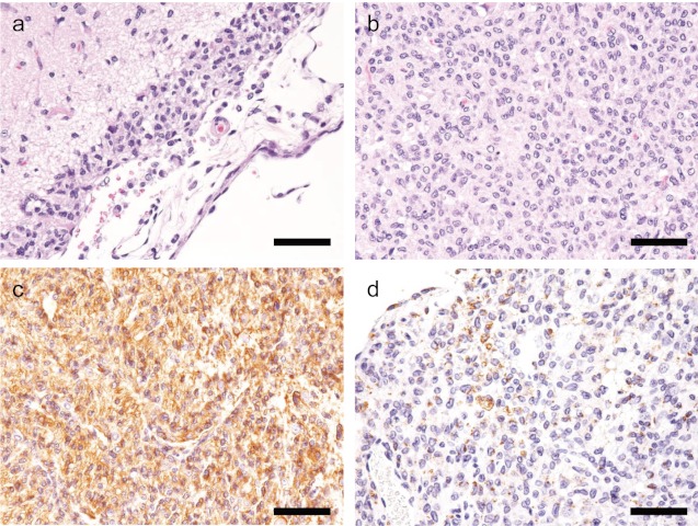
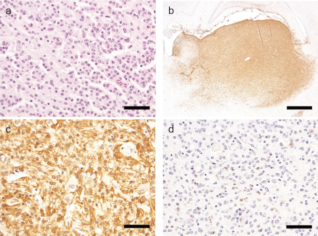
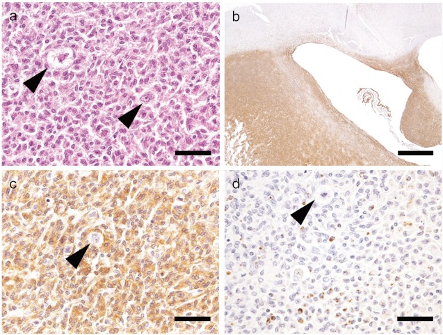
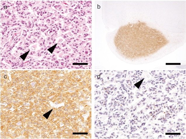
In rats, it is sometimes difficult to distinguish malignant reticuloses from astrocytomas in routine histopathological assessment. In the present study, four spontaneous brain neoplasms developing in the cerebrum of one Wistar Hannover rat and three Sprague-Dawley rats were immunohistochemically examined using microglia and macrophage markers. Histopathologically, these neoplasms were localized mainly in the cerebral cortex, hypothalamus or piriform lobe, and the portions showing solid growth did not show characteristic cellular arrangement but had an indistinct boundary with the surrounding brain parenchyma. Neoplastic cells had oval or pleomorphic small nuclei with abundant eosinophilic cytoplasm. Two cases showed neoplastic cell infiltration into the meninges and perivascular spaces. Silver staining showed lack of reticulin fiber production in the stroma of the neoplasms. Immunohistochemically, the neoplastic cells were strongly positive for Iba-1 and sporadically positive for CD68 in all four cases. On the basis of these results, all the neoplasms examined here could be distinguished from astrocytomas and diagnosed as malignant reticuloses. Thus, immunohistochemical demonstration of microglia/macrophage characters, such as using Iba-1, is considered to be helpful for differential diagnosis of malignant reticuloses from astrocytomas among spontaneously occurring primary brain neoplasms in rats.
在大鼠中,在常规组织病理学评估中有时很难区分恶性网状细胞增生症和星形细胞瘤。在本研究中,对一只Wistar Hannover大鼠和三只Sprague-Dawley大鼠大脑中发生的四个自发性脑肿瘤进行了免疫组织化学检查,使用了小胶质细胞和巨噬细胞标志物。组织病理学上,这些肿瘤主要位于大脑皮层、下丘脑或梨状叶,呈实体生长的部分没有特征性的细胞排列,但与周围脑实质的边界不清晰。肿瘤细胞有椭圆形或多形性的小核,胞质嗜酸性丰富。两例显示肿瘤细胞浸润到脑膜和血管周围间隙。银染色显示肿瘤间质中缺乏网状纤维产生。免疫组织化学上,所有四例肿瘤细胞对Iba-1均呈强阳性,对CD68呈散在阳性。基于这些结果,此处检查的所有肿瘤均可与星形细胞瘤区分开来,并诊断为恶性网状细胞增生症。因此,免疫组织化学显示小胶质细胞/巨噬细胞特征,如使用Iba-1,被认为有助于在大鼠自发性原发性脑肿瘤中鉴别恶性网状细胞增生症和星形细胞瘤。