Laboratory of Environmental Toxicology, National Institute of Chemical Physics and Biophysics, Tallinn, Estonia.
PLoS One. 2013 May 30;8(5):e64060. doi: 10.1371/journal.pone.0064060. Print 2013.
It is generally accepted that antibacterial properties of Ag nanoparticles (AgNPs) are dictated by their dissolved fraction. However, dissolution-based concept alone does not fully explain the toxic potency of nanoparticulate silver compared to silver ions.
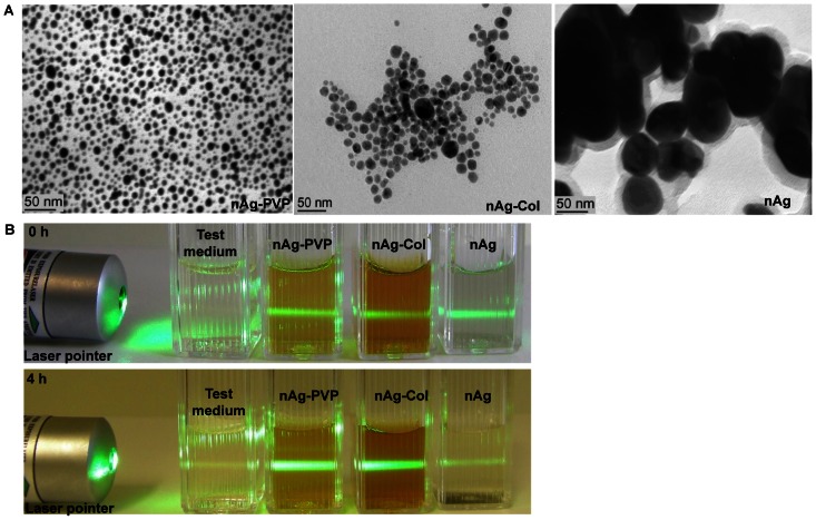
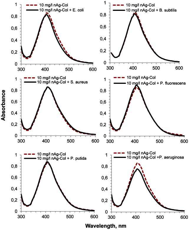
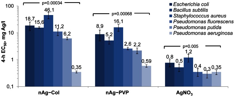
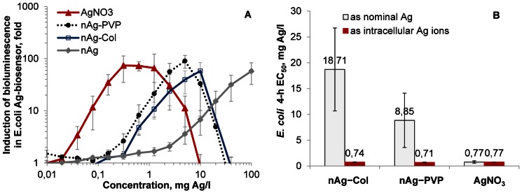
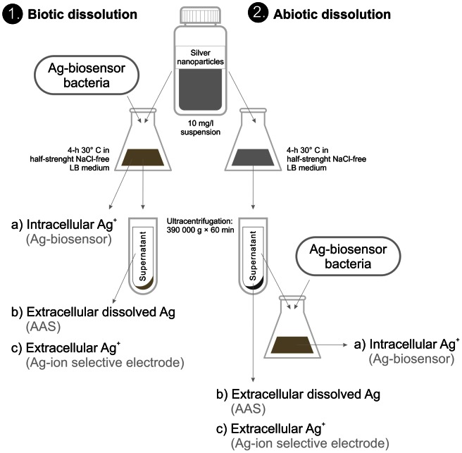
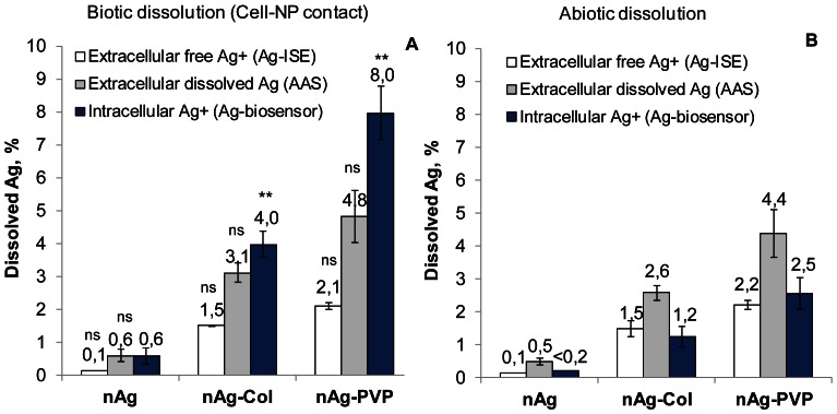
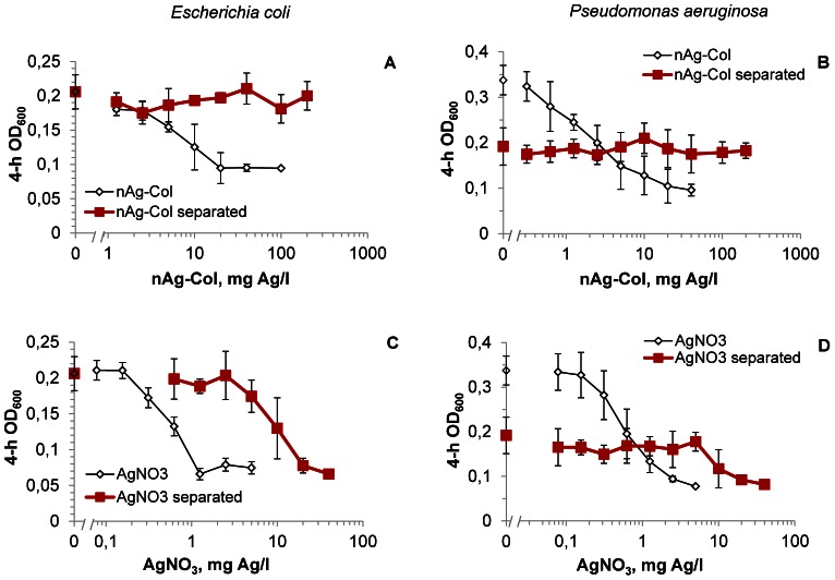
METHODOLOGY/PRINCIPAL FINDINGS: Herein, we demonstrated that the direct contact between bacterial cell and AgNPs' surface enhanced the toxicity of nanosilver. More specifically, cell-NP contact increased the cellular uptake of particle-associated Ag ions - the single and ultimate cause of toxicity. To prove that, we evaluated the toxicity of three different AgNPs (uncoated, PVP-coated and protein-coated) to six bacterial strains: Gram-negative Escherichia coli, Pseudomonas fluorescens, P. putida and P. aeruginosa and Gram-positive Bacillus subtilis and Staphylococcus aureus. While the toxicity of AgNO3 to these bacteria varied only slightly (the 4-h EC50 ranged from 0.3 to 1.2 mg Ag/l), the 4-h EC50 values of protein-coated AgNPs for various bacterial strains differed remarkably, from 0.35 to 46 mg Ag/l. By systematically comparing the intracellular and extracellular free Ag(+) liberated from AgNPs, we demonstrated that not only extracellular dissolution in the bacterial test environment but also additional dissolution taking place at the particle-cell interface played an essential role in antibacterial action of AgNPs. The role of the NP-cell contact in dictating the antibacterial activity of Ag-NPs was additionally proven by the following observations: (i) separation of bacterial cells from AgNPs by particle-impermeable membrane (cut-off 20 kDa, ∼4 nm) significantly reduced the toxicity of AgNPs and (ii) P. aeruginosa cells which tended to attach onto AgNPs, exhibited the highest sensitivity to all forms of nanoparticulate Ag.
CONCLUSIONS/SIGNIFICANCE: Our findings provide new insights into the mode of antibacterial action of nanosilver and explain some discrepancies in this field, showing that "Ag-ion" and "particle-specific" mechanisms are not controversial but, rather, are two faces of the same coin.
人们普遍认为,银纳米粒子(AgNPs)的抗菌性能取决于其溶解部分。然而,仅基于溶解的概念并不能完全解释纳米银相对于银离子的毒性强度。
方法/主要发现:本文证明了细菌细胞与 AgNPs 表面的直接接触增强了纳米银的毒性。具体而言,细胞与 NP 的接触增加了颗粒相关银离子的细胞内摄取——这是毒性的单一且最终原因。为了证明这一点,我们评估了三种不同的 AgNPs(未涂层、PVP 涂层和蛋白质涂层)对六种细菌菌株的毒性:革兰氏阴性大肠杆菌、荧光假单胞菌、铜绿假单胞菌和鲍曼不动杆菌以及革兰氏阳性枯草芽孢杆菌和金黄色葡萄球菌。虽然 AgNO3 对这些细菌的毒性差异很小(4 小时 EC50 范围为 0.3 至 1.2 mg Ag/l),但各种细菌菌株的蛋白质涂层 AgNPs 的 4 小时 EC50 值差异很大,从 0.35 至 46 mg Ag/l。通过系统比较从 AgNPs 中释放的细胞内和细胞外游离的 Ag(+),我们证明,不仅是细菌测试环境中的细胞外溶解,而且在颗粒-细胞界面发生的额外溶解在 AgNPs 的抗菌作用中发挥了重要作用。Ag-NPs 的抗菌活性取决于 NP-细胞接触的作用,这一点还可以通过以下观察结果得到证明:(i)通过粒子不可渗透的膜(截留 20 kDa,约 4 nm)将细菌细胞与 AgNPs 分离,显著降低了 AgNPs 的毒性;(ii)倾向于附着在 AgNPs 上的铜绿假单胞菌细胞对所有形式的纳米银都表现出最高的敏感性。
结论/意义:我们的发现为纳米银的抗菌作用模式提供了新的见解,并解释了该领域的一些差异,表明“Ag 离子”和“颗粒特异性”机制不是相互矛盾的,而是同一枚硬币的两面。