• 文献检索
  • 文档翻译
  • 深度研究
  • 学术资讯
  • Suppr Zotero 插件Zotero 插件
  • 邀请有礼
  • 套餐&价格
  • 历史记录
应用&插件
Suppr Zotero 插件Zotero 插件浏览器插件Mac 客户端Windows 客户端微信小程序
定价
高级版会员购买积分包购买API积分包
服务
文献检索文档翻译深度研究API 文档MCP 服务
关于我们
关于 Suppr公司介绍联系我们用户协议隐私条款
关注我们

Suppr 超能文献

核心技术专利:CN118964589B侵权必究
粤ICP备2023148730 号-1Suppr @ 2026

文献检索

告别复杂PubMed语法,用中文像聊天一样搜索,搜遍4000万医学文献。AI智能推荐,让科研检索更轻松。

立即免费搜索

文件翻译

保留排版,准确专业,支持PDF/Word/PPT等文件格式,支持 12+语言互译。

免费翻译文档

深度研究

AI帮你快速写综述,25分钟生成高质量综述,智能提取关键信息,辅助科研写作。

立即免费体验

来自人类蛋白质图谱的乳腺组织免疫染色模式的自动分类

Automated classification of immunostaining patterns in breast tissue from the human protein atlas.

作者信息

Swamidoss Issac Niwas, Kårsnäs Andreas, Uhlmann Virginie, Ponnusamy Palanisamy, Kampf Caroline, Simonsson Martin, Wählby Carolina, Strand Robin

机构信息

Department of Electronics and Communication Engineering, National Institute of Technology (NIT), Tiruchirappalli, Tamil Nadu, India ; Centre for Image Analysis (CBA) and SciLife Lab, Uppsala University, Uppsala, Sweden.

出版信息

J Pathol Inform. 2013 Mar 30;4(Suppl):S14. doi: 10.4103/2153-3539.109881. Print 2013.

DOI:10.4103/2153-3539.109881
PMID:23766936
原文链接:https://pmc.ncbi.nlm.nih.gov/articles/PMC3678740/
Abstract

BACKGROUND

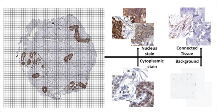
The Human Protein Atlas (HPA) is an effort to map the location of all human proteins (http://www.proteinatlas.org/). It contains a large number of histological images of sections from human tissue. Tissue micro arrays (TMA) are imaged by a slide scanning microscope, and each image represents a thin slice of a tissue core with a dark brown antibody specific stain and a blue counter stain. When generating antibodies for protein profiling of the human proteome, an important step in the quality control is to compare staining patterns of different antibodies directed towards the same protein. This comparison is an ultimate control that the antibody recognizes the right protein. In this paper, we propose and evaluate different approaches for classifying sub-cellular antibody staining patterns in breast tissue samples.

MATERIALS AND METHODS

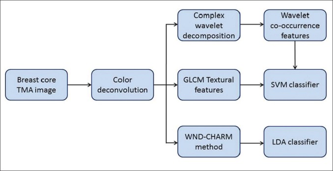
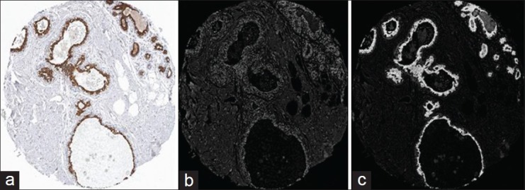
The proposed methods include the computation of various features including gray level co-occurrence matrix (GLCM) features, complex wavelet co-occurrence matrix (CWCM) features, and weighted neighbor distance using compound hierarchy of algorithms representing morphology (WND-CHARM)-inspired features. The extracted features are used into two different multivariate classifiers (support vector machine (SVM) and linear discriminant analysis (LDA) classifier). Before extracting features, we use color deconvolution to separate different tissue components, such as the brownly stained positive regions and the blue cellular regions, in the immuno-stained TMA images of breast tissue.

RESULTS

We present classification results based on combinations of feature measurements. The proposed complex wavelet features and the WND-CHARM features have accuracy similar to that of a human expert.

CONCLUSIONS

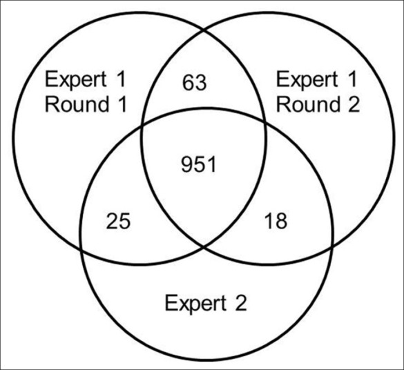
Both human experts and the proposed automated methods have difficulties discriminating between nuclear and cytoplasmic staining patterns. This is to a large extent due to mixed staining of nucleus and cytoplasm. Methods for quantification of staining patterns in histopathology have many applications, ranging from antibody quality control to tumor grading.

摘要

背景

人类蛋白质图谱(HPA)致力于绘制所有人类蛋白质的位置(http://www.proteinatlas.org/)。它包含大量人体组织切片的组织学图像。组织微阵列(TMA)通过载玻片扫描显微镜成像,每张图像代表一个组织芯的薄片,带有深棕色抗体特异性染色和蓝色复染。在生成用于人类蛋白质组蛋白质谱分析的抗体时,质量控制中的一个重要步骤是比较针对同一蛋白质的不同抗体的染色模式。这种比较是对抗体识别正确蛋白质的最终控制。在本文中,我们提出并评估了用于对乳腺组织样本中的亚细胞抗体染色模式进行分类的不同方法。

材料与方法

所提出的方法包括计算各种特征,包括灰度共生矩阵(GLCM)特征、复小波共生矩阵(CWCM)特征以及使用表示形态学的算法复合层次结构的加权邻域距离(WND-CHARM)启发的特征。提取的特征被用于两种不同的多变量分类器(支持向量机(SVM)和线性判别分析(LDA)分类器)。在提取特征之前,我们使用颜色反卷积来分离乳腺组织免疫染色TMA图像中的不同组织成分,如棕色染色的阳性区域和蓝色细胞区域。

结果

我们展示了基于特征测量组合的分类结果。所提出的复小波特征和WND-CHARM特征的准确性与人类专家相似。

结论

人类专家和所提出的自动化方法在区分核染色模式和细胞质染色模式方面都存在困难。这在很大程度上是由于细胞核和细胞质的混合染色。组织病理学中染色模式定量方法有许多应用,从抗体质量控制到肿瘤分级。

https://cdn.ncbi.nlm.nih.gov/pmc/blobs/5adc/3678740/cab2eb308c71/JPI-4-14-g004.jpg
https://cdn.ncbi.nlm.nih.gov/pmc/blobs/5adc/3678740/aaf9218515cc/JPI-4-14-g001.jpg
https://cdn.ncbi.nlm.nih.gov/pmc/blobs/5adc/3678740/6dec361c6b08/JPI-4-14-g002.jpg
https://cdn.ncbi.nlm.nih.gov/pmc/blobs/5adc/3678740/ff29069f347c/JPI-4-14-g003.jpg
https://cdn.ncbi.nlm.nih.gov/pmc/blobs/5adc/3678740/cab2eb308c71/JPI-4-14-g004.jpg

相似文献

1
Automated classification of immunostaining patterns in breast tissue from the human protein atlas.来自人类蛋白质图谱的乳腺组织免疫染色模式的自动分类
J Pathol Inform. 2013 Mar 30;4(Suppl):S14. doi: 10.4103/2153-3539.109881. Print 2013.
2
Computer-assisted lip diagnosis on Traditional Chinese Medicine using multi-class support vector machines.基于多类支持向量机的中医唇诊计算机辅助诊断。
BMC Complement Altern Med. 2012 Aug 16;12:127. doi: 10.1186/1472-6882-12-127.
3
Appearance and characterization of fruit image textures for quality sorting using wavelet transform and genetic algorithms.利用小波变换和遗传算法对水果图像纹理进行外观和特征描述,以实现品质分选。
J Texture Stud. 2018 Feb;49(1):65-83. doi: 10.1111/jtxs.12284. Epub 2017 Aug 6.
4
Mammographic masses characterization based on localized texture and dataset fractal analysis using linear, neural and support vector machine classifiers.基于局部纹理和数据集分形分析,使用线性、神经网络和支持向量机分类器对乳腺X线摄影肿块进行特征描述。
Artif Intell Med. 2006 Jun;37(2):145-62. doi: 10.1016/j.artmed.2006.03.002. Epub 2006 May 23.
5
Phenotype recognition with combined features and random subspace classifier ensemble.基于组合特征和随机子空间分类器集成的表型识别。
BMC Bioinformatics. 2011 Apr 30;12:128. doi: 10.1186/1471-2105-12-128.
6
AutoIHC-Analyzer: computer-assisted microscopy for automated membrane extraction/scoring in HER2 molecular markers.AutoIHC-Analyzer:用于 HER2 分子标志物的自动膜提取/评分的计算机辅助显微镜。
J Microsc. 2021 Jan;281(1):87-96. doi: 10.1111/jmi.12955. Epub 2020 Aug 27.
7
Differentiation of fat-poor angiomyolipoma from clear cell renal cell carcinoma in contrast-enhanced MDCT images using quantitative feature classification.基于定量特征分类的 MDCT 增强图像鉴别乏脂性血管平滑肌脂肪瘤与透明细胞肾细胞癌
Med Phys. 2017 Jul;44(7):3604-3614. doi: 10.1002/mp.12258. Epub 2017 Jun 9.
8
Computer-aided diagnosis system for classifying benign and malignant thyroid nodules in multi-stained FNAB cytological images.用于在多染色细针穿刺抽吸活检(FNAB)细胞学图像中对甲状腺良恶性结节进行分类的计算机辅助诊断系统。
Australas Phys Eng Sci Med. 2013 Jun;36(2):219-30. doi: 10.1007/s13246-013-0199-8. Epub 2013 May 21.
9
Automated discrimination of lower and higher grade gliomas based on histopathological image analysis.基于组织病理学图像分析的低级别和高级别胶质瘤自动鉴别
J Pathol Inform. 2015 Mar 24;6:15. doi: 10.4103/2153-3539.153914. eCollection 2015.
10
Full Intelligent Cancer Classification of Thermal Breast Images to Assist Physician in Clinical Diagnostic Applications.用于临床诊断应用中辅助医生的乳腺热图像全智能癌症分类
J Med Signals Sens. 2016 Jan-Mar;6(1):12-24.

引用本文的文献

1
Serine protease PRSS23 drives gastric cancer by enhancing tumor associated macrophage infiltration FGF2.丝氨酸蛋白酶 PRSS23 通过增强肿瘤相关巨噬细胞浸润 FGF2 促进胃癌发生。
Front Immunol. 2022 Sep 15;13:955841. doi: 10.3389/fimmu.2022.955841. eCollection 2022.
2
IRF6 Is Directly Regulated by ZEB1 and ELF3, and Predicts a Favorable Prognosis in Gastric Cancer.IRF6受ZEB1和ELF3直接调控,并预测胃癌的预后良好。
Front Oncol. 2019 Apr 4;9:220. doi: 10.3389/fonc.2019.00220. eCollection 2019.
3
The Need for Careful Data Collection for Pattern Recognition in Digital Pathology.

本文引用的文献

1
ImmunoMembrane: a publicly available web application for digital image analysis of HER2 immunohistochemistry.ImmunoMembrane:一个公开的可用的用于 HER2 免疫组化数字图像分析的网络应用程序。
Histopathology. 2012 Apr;60(5):758-67. doi: 10.1111/j.1365-2559.2011.04142.x. Epub 2012 Feb 1.
2
Assessing the efficacy of low-level image content descriptors for computer-based fluorescence microscopy image analysis.评估基于计算机的荧光显微镜图像分析中低水平图像内容描述符的功效。
J Microsc. 2011 Sep;243(3):284-92. doi: 10.1111/j.1365-2818.2011.03502.x. Epub 2011 May 23.
3
ImmunoRatio: a publicly available web application for quantitative image analysis of estrogen receptor (ER), progesterone receptor (PR), and Ki-67.
数字病理学中模式识别所需的仔细数据收集
J Pathol Inform. 2017 Apr 10;8:19. doi: 10.4103/jpi.jpi_94_16. eCollection 2017.
4
Proteomics in India: the clinical aspect.印度的蛋白质组学:临床方面。
Clin Proteomics. 2016 Nov 5;13:21. doi: 10.1186/s12014-016-9122-0. eCollection 2016.
5
CP-CHARM: segmentation-free image classification made accessible.CP-CHARM:实现无分割图像分类。
BMC Bioinformatics. 2016 Jan 27;17:51. doi: 10.1186/s12859-016-0895-y.
6
The influence of the microscope lamp filament colour temperature on the process of digital images of histological slides acquisition standardization.显微镜灯光丝色温对组织学切片数字图像采集标准化过程的影响。
Diagn Pathol. 2014;9 Suppl 1(Suppl 1):S13. doi: 10.1186/1746-1596-9-S1-S13. Epub 2014 Dec 19.
7
Image segmentation and identification of paired antibodies in breast tissue.乳腺组织中配对抗体的图像分割与识别。
Comput Math Methods Med. 2014;2014:647273. doi: 10.1155/2014/647273. Epub 2014 Jul 1.
8
Expression and prognostic significance of the polymeric immunoglobulin receptor in epithelial ovarian cancer.聚合免疫球蛋白受体在上皮性卵巢癌中的表达及预后意义
J Ovarian Res. 2014 Feb 26;7:26. doi: 10.1186/1757-2215-7-26.
ImmunoRatio:一个可公开获取的用于雌激素受体(ER)、孕激素受体(PR)和 Ki-67 定量图像分析的网络应用程序。
Breast Cancer Res. 2010;12(4):R56. doi: 10.1186/bcr2615. Epub 2010 Jul 27.
4
Statistical nuclear texture analysis in cancer research: a review of methods and applications.癌症研究中的统计性核纹理分析:方法与应用综述
Crit Rev Oncog. 2008;14(2-3):89-164. doi: 10.1615/critrevoncog.v14.i2-3.10.
5
WND-CHARM: Multi-purpose image classification using compound image transforms.WND-CHARM:使用复合图像变换的多用途图像分类
Pattern Recognit Lett. 2008 Jan;29(11):1684-1693. doi: 10.1016/j.patrec.2008.04.013.
6
Novel image analysis approach for quantifying expression of nuclear proteins assessed by immunohistochemistry: application to measurement of oestrogen and progesterone receptor levels in breast cancer.用于通过免疫组织化学定量评估核蛋白表达的新型图像分析方法:在乳腺癌雌激素和孕激素受体水平测量中的应用。
Breast Cancer Res. 2008;10(5):R89. doi: 10.1186/bcr2187. Epub 2008 Oct 23.
7
Wndchrm - an open source utility for biological image analysis.Wndchrm - 一款用于生物图像分析的开源实用工具。
Source Code Biol Med. 2008 Jul 8;3:13. doi: 10.1186/1751-0473-3-13.
8
A framework for the automated analysis of subcellular patterns in human protein atlas images.人类蛋白质图谱图像中亚细胞模式自动分析框架。
J Proteome Res. 2008 Jun;7(6):2300-8. doi: 10.1021/pr7007626. Epub 2008 Apr 25.
9
CellProfiler: free, versatile software for automated biological image analysis.细胞图像分析软件(CellProfiler):用于自动生物图像分析的免费通用软件。
Biotechniques. 2007 Jan;42(1):71-5. doi: 10.2144/000112257.
10
Quantification of immunohistochemistry--issues concerning methods, utility and semiquantitative assessment II.免疫组织化学的定量分析——关于方法、实用性及半定量评估的问题II
Histopathology. 2006 Oct;49(4):411-24. doi: 10.1111/j.1365-2559.2006.02513.x.