• 文献检索
  • 文档翻译
  • 深度研究
  • 学术资讯
  • Suppr Zotero 插件Zotero 插件
  • 邀请有礼
  • 套餐&价格
  • 历史记录
应用&插件
Suppr Zotero 插件Zotero 插件浏览器插件Mac 客户端Windows 客户端微信小程序
定价
高级版会员购买积分包购买API积分包
服务
文献检索文档翻译深度研究API 文档MCP 服务
关于我们
关于 Suppr公司介绍联系我们用户协议隐私条款
关注我们

Suppr 超能文献

核心技术专利:CN118964589B侵权必究
粤ICP备2023148730 号-1Suppr @ 2026

文献检索

告别复杂PubMed语法,用中文像聊天一样搜索,搜遍4000万医学文献。AI智能推荐,让科研检索更轻松。

立即免费搜索

文件翻译

保留排版,准确专业,支持PDF/Word/PPT等文件格式,支持 12+语言互译。

免费翻译文档

深度研究

AI帮你快速写综述,25分钟生成高质量综述,智能提取关键信息,辅助科研写作。

立即免费体验

CP-CHARM:实现无分割图像分类。

CP-CHARM: segmentation-free image classification made accessible.

作者信息

Uhlmann Virginie, Singh Shantanu, Carpenter Anne E

机构信息

Biomedical Imaging Group, Swiss Federal Institute of Technology (EPFL), Lausanne, Switzerland.

Imaging Platform, Broad Institute of Harvard and MIT, Cambridge, MA, USA.

出版信息

BMC Bioinformatics. 2016 Jan 27;17:51. doi: 10.1186/s12859-016-0895-y.

DOI:10.1186/s12859-016-0895-y
PMID:26817459
原文链接:https://pmc.ncbi.nlm.nih.gov/articles/PMC4729047/
Abstract

BACKGROUND

Automated classification using machine learning often relies on features derived from segmenting individual objects, which can be difficult to automate. WND-CHARM is a previously developed classification algorithm in which features are computed on the whole image, thereby avoiding the need for segmentation. The algorithm obtained encouraging results but requires considerable computational expertise to execute. Furthermore, some benchmark sets have been shown to be subject to confounding artifacts that overestimate classification accuracy.

RESULTS

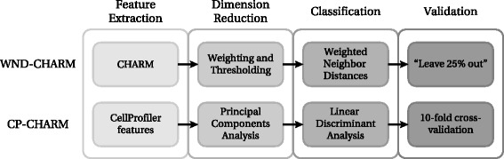
We developed CP-CHARM, a user-friendly image-based classification algorithm inspired by WND-CHARM in (i) its ability to capture a wide variety of morphological aspects of the image, and (ii) the absence of requirement for segmentation. In order to make such an image-based classification method easily accessible to the biological research community, CP-CHARM relies on the widely-used open-source image analysis software CellProfiler for feature extraction. To validate our method, we reproduced WND-CHARM's results and ensured that CP-CHARM obtained comparable performance. We then successfully applied our approach on cell-based assay data and on tissue images. We designed these new training and test sets to reduce the effect of batch-related artifacts.

CONCLUSIONS

The proposed method preserves the strengths of WND-CHARM - it extracts a wide variety of morphological features directly on whole images thereby avoiding the need for cell segmentation, but additionally, it makes the methods easily accessible for researchers without computational expertise by implementing them as a CellProfiler pipeline. It has been demonstrated to perform well on a wide range of bioimage classification problems, including on new datasets that have been carefully selected and annotated to minimize batch effects. This provides for the first time a realistic and reliable assessment of the whole image classification strategy.

摘要

背景

使用机器学习的自动分类通常依赖于从单个对象分割中派生的特征,而这可能难以实现自动化。WND-CHARM是一种先前开发的分类算法,其特征是在整个图像上计算的,从而避免了分割的需要。该算法取得了令人鼓舞的结果,但需要相当多的计算专业知识才能执行。此外,一些基准集已被证明存在混淆伪影,会高估分类准确性。

结果

我们开发了CP-CHARM,这是一种用户友好的基于图像的分类算法,其灵感来自于WND-CHARM的以下两点:(i)捕捉图像各种形态学方面的能力,以及(ii)无需分割的特点。为了使生物研究界能够轻松使用这种基于图像的分类方法,CP-CHARM依赖于广泛使用的开源图像分析软件CellProfiler进行特征提取。为了验证我们的方法,我们重现了WND-CHARM的结果,并确保CP-CHARM获得了可比的性能。然后,我们成功地将我们的方法应用于基于细胞的检测数据和组织图像。我们设计了这些新的训练集和测试集,以减少批次相关伪影的影响。

结论

所提出的方法保留了WND-CHARM的优点——它直接在整个图像上提取各种形态特征,从而避免了细胞分割的需要,但此外,通过将其实现为CellProfiler管道,它使没有计算专业知识的研究人员也能轻松使用这些方法。已证明它在广泛的生物图像分类问题上表现良好,包括在经过精心挑选和注释以最小化批次效应的新数据集上。这首次为全图像分类策略提供了现实可靠的评估。

https://cdn.ncbi.nlm.nih.gov/pmc/blobs/dce7/4729047/4c26b9efc196/12859_2016_895_Fig3_HTML.jpg
https://cdn.ncbi.nlm.nih.gov/pmc/blobs/dce7/4729047/6b372aef899d/12859_2016_895_Fig1_HTML.jpg
https://cdn.ncbi.nlm.nih.gov/pmc/blobs/dce7/4729047/c421aa048f34/12859_2016_895_Fig2_HTML.jpg
https://cdn.ncbi.nlm.nih.gov/pmc/blobs/dce7/4729047/4c26b9efc196/12859_2016_895_Fig3_HTML.jpg
https://cdn.ncbi.nlm.nih.gov/pmc/blobs/dce7/4729047/6b372aef899d/12859_2016_895_Fig1_HTML.jpg
https://cdn.ncbi.nlm.nih.gov/pmc/blobs/dce7/4729047/c421aa048f34/12859_2016_895_Fig2_HTML.jpg
https://cdn.ncbi.nlm.nih.gov/pmc/blobs/dce7/4729047/4c26b9efc196/12859_2016_895_Fig3_HTML.jpg

相似文献

1
CP-CHARM: segmentation-free image classification made accessible.CP-CHARM:实现无分割图像分类。
BMC Bioinformatics. 2016 Jan 27;17:51. doi: 10.1186/s12859-016-0895-y.
2
FogBank: a single cell segmentation across multiple cell lines and image modalities.FogBank:跨多种细胞系和图像模态的单细胞分割
BMC Bioinformatics. 2014 Dec 30;15(1):431. doi: 10.1186/s12859-014-0431-x.
3
Phenotype recognition with combined features and random subspace classifier ensemble.基于组合特征和随机子空间分类器集成的表型识别。
BMC Bioinformatics. 2011 Apr 30;12:128. doi: 10.1186/1471-2105-12-128.
4
Robust and automated three-dimensional segmentation of densely packed cell nuclei in different biological specimens with Lines-of-Sight decomposition.利用视线分解对不同生物样本中紧密堆积的细胞核进行稳健且自动化的三维分割。
BMC Bioinformatics. 2015 Jun 8;16:187. doi: 10.1186/s12859-015-0617-x.
5
Cell segmentation in phase contrast microscopy images via semi-supervised classification over optics-related features.基于与光学相关特征的半监督分类的相差显微镜图像中的细胞分割。
Med Image Anal. 2013 Oct;17(7):746-65. doi: 10.1016/j.media.2013.04.004. Epub 2013 Apr 29.
6
WND-CHARM: Multi-purpose image classification using compound image transforms.WND-CHARM:使用复合图像变换的多用途图像分类
Pattern Recognit Lett. 2008 Jan;29(11):1684-1693. doi: 10.1016/j.patrec.2008.04.013.
7
Segmentation of cDNA microarray spots using markov random field modeling.使用马尔可夫随机场建模对cDNA微阵列斑点进行分割。
Bioinformatics. 2005 Jul 1;21(13):2994-3000. doi: 10.1093/bioinformatics/bti455. Epub 2005 Apr 19.
8
A multichannel watershed-based segmentation method for multispectral chromosome classification.一种基于多通道分水岭算法的多光谱染色体分类分割方法。
IEEE Trans Med Imaging. 2008 May;27(5):697-708. doi: 10.1109/TMI.2008.916962.
9
Donuts, scratches and blanks: robust model-based segmentation of microarray images.圆环、划痕与空白:基于稳健模型的微阵列图像分割
Bioinformatics. 2005 Jun 15;21(12):2875-82. doi: 10.1093/bioinformatics/bti447. Epub 2005 Apr 21.
10
BIOCAT: a pattern recognition platform for customizable biological image classification and annotation.BIOCAT:一个用于可定制生物图像分类和标注的模式识别平台。
BMC Bioinformatics. 2013 Oct 4;14:291. doi: 10.1186/1471-2105-14-291.

引用本文的文献

1
Unbiased identification of cell identity in dense mixed neural cultures.在密集混合神经培养物中无偏倚地识别细胞身份。
Elife. 2025 Jan 17;13:RP95273. doi: 10.7554/eLife.95273.
2
TDAExplore: Quantitative analysis of fluorescence microscopy images through topology-based machine learning.TDAExplore:通过基于拓扑学的机器学习对荧光显微镜图像进行定量分析。
Patterns (N Y). 2021 Oct 12;2(11):100367. doi: 10.1016/j.patter.2021.100367. eCollection 2021 Nov 12.
3
Rapid 3D phenotypic analysis of neurons and organoids using data-driven cell segmentation-free machine learning.

本文引用的文献

1
Review of free software tools for image analysis of fluorescence cell micrographs.用于荧光细胞显微图像分析的免费软件工具综述。
J Microsc. 2015 Jan;257(1):39-53. doi: 10.1111/jmi.12184. Epub 2014 Oct 31.
2
Many local pattern texture features: which is better for image-based multilabel human protein subcellular localization classification?多种局部模式纹理特征:哪种特征更适合基于图像的多标签人类蛋白质亚细胞定位分类?
ScientificWorldJournal. 2014;2014:429049. doi: 10.1155/2014/429049. Epub 2014 Jun 24.
3
BIOCAT: a pattern recognition platform for customizable biological image classification and annotation.
使用基于数据驱动的无细胞分割机器学习技术快速进行神经元和类器官的 3D 表型分析。
PLoS Comput Biol. 2021 Feb 22;17(2):e1008630. doi: 10.1371/journal.pcbi.1008630. eCollection 2021 Feb.
4
Domain-invariant features for mechanism of action prediction in a multi-cell-line drug screen.用于多细胞系药物筛选中作用机制预测的域不变特征。
Bioinformatics. 2020 Mar 1;36(5):1607-1613. doi: 10.1093/bioinformatics/btz774.
5
Automated and Reproducible Detection of Vascular Endothelial Growth Factor (VEGF) in Renal Tissue Sections.自动且可重现的肾组织切片中血管内皮生长因子(VEGF)的检测。
J Immunol Res. 2019 Mar 19;2019:7232781. doi: 10.1155/2019/7232781. eCollection 2019.
6
Transfer Learning with Deep Convolutional Neural Networks for Classifying Cellular Morphological Changes.基于深度卷积神经网络的迁移学习在细胞形态变化分类中的应用
SLAS Discov. 2019 Apr;24(4):466-475. doi: 10.1177/2472555218818756. Epub 2019 Jan 14.
7
Automated Neuron Detection in High-Content Fluorescence Microscopy Images Using Machine Learning.基于机器学习的高内涵荧光显微镜图像中的自动神经元检测。
Neuroinformatics. 2019 Apr;17(2):253-269. doi: 10.1007/s12021-018-9399-4.
8
Machine learning and image-based profiling in drug discovery.药物研发中的机器学习与基于图像的分析
Curr Opin Syst Biol. 2018 Aug;10:43-52. doi: 10.1016/j.coisb.2018.05.004.
9
Environmental properties of cells improve machine learning-based phenotype recognition accuracy.细胞的环境特性提高了基于机器学习的表型识别准确性。
Sci Rep. 2018 Jul 4;8(1):10085. doi: 10.1038/s41598-018-28482-y.
10
Sharing and reusing cell image data.细胞图像数据的共享与再利用。
Mol Biol Cell. 2018 Jun 1;29(11):1274-1280. doi: 10.1091/mbc.E17-10-0606.
BIOCAT:一个用于可定制生物图像分类和标注的模式识别平台。
BMC Bioinformatics. 2013 Oct 4;14:291. doi: 10.1186/1471-2105-14-291.
4
Determining the subcellular location of new proteins from microscope images using local features.使用局部特征从显微镜图像中确定新蛋白质的亚细胞位置。
Bioinformatics. 2013 Sep 15;29(18):2343-9. doi: 10.1093/bioinformatics/btt392. Epub 2013 Jul 8.
5
Automated classification of immunostaining patterns in breast tissue from the human protein atlas.来自人类蛋白质图谱的乳腺组织免疫染色模式的自动分类
J Pathol Inform. 2013 Mar 30;4(Suppl):S14. doi: 10.4103/2153-3539.109881. Print 2013.
6
Annotated high-throughput microscopy image sets for validation.用于验证的带注释的高通量显微镜图像集。
Nat Methods. 2012 Jun 28;9(7):637. doi: 10.1038/nmeth.2083.
7
PhenoRipper: software for rapidly profiling microscopy images.PhenoRipper:用于快速分析显微镜图像的软件。
Nat Methods. 2012 Jun 28;9(7):635-7. doi: 10.1038/nmeth.2097.
8
Assessing the efficacy of low-level image content descriptors for computer-based fluorescence microscopy image analysis.评估基于计算机的荧光显微镜图像分析中低水平图像内容描述符的功效。
J Microsc. 2011 Sep;243(3):284-92. doi: 10.1111/j.1365-2818.2011.03502.x. Epub 2011 May 23.
9
Improved structure, function and compatibility for CellProfiler: modular high-throughput image analysis software.改进 CellProfiler 的结构、功能和兼容性:用于高通量图像分析的模块化软件。
Bioinformatics. 2011 Apr 15;27(8):1179-80. doi: 10.1093/bioinformatics/btr095. Epub 2011 Feb 23.
10
Towards a knowledge-based Human Protein Atlas.迈向基于知识的人类蛋白质图谱。
Nat Biotechnol. 2010 Dec;28(12):1248-50. doi: 10.1038/nbt1210-1248.