• 文献检索
  • 文档翻译
  • 深度研究
  • 学术资讯
  • Suppr Zotero 插件Zotero 插件
  • 邀请有礼
  • 套餐&价格
  • 历史记录
应用&插件
Suppr Zotero 插件Zotero 插件浏览器插件Mac 客户端Windows 客户端微信小程序
定价
高级版会员购买积分包购买API积分包
服务
文献检索文档翻译深度研究API 文档MCP 服务
关于我们
关于 Suppr公司介绍联系我们用户协议隐私条款
关注我们

Suppr 超能文献

核心技术专利:CN118964589B侵权必究
粤ICP备2023148730 号-1Suppr @ 2026

文献检索

告别复杂PubMed语法,用中文像聊天一样搜索,搜遍4000万医学文献。AI智能推荐,让科研检索更轻松。

立即免费搜索

文件翻译

保留排版,准确专业,支持PDF/Word/PPT等文件格式,支持 12+语言互译。

免费翻译文档

深度研究

AI帮你快速写综述,25分钟生成高质量综述,智能提取关键信息,辅助科研写作。

立即免费体验

大孔结构增强静电纺丝支架的血管化。

Macroporosity enhances vascularization of electrospun scaffolds.

机构信息

Department of Surgery, University of California, Los Angeles, California 90095, USA.

出版信息

J Surg Res. 2013 Jul;183(1):18-26. doi: 10.1016/j.jss.2013.01.005. Epub 2013 Feb 1.

DOI:10.1016/j.jss.2013.01.005
PMID:23769018
原文链接:https://pmc.ncbi.nlm.nih.gov/articles/PMC3694756/
Abstract

BACKGROUND

One of the greatest challenges in scaffold-based tissue engineering remains poor and inefficient penetration of cells into scaffolds to generate thick vascularized and cellular tissues. Electrospinning has emerged as a preferred method for producing scaffolds with high surface area-to-volume ratios and resemblance to extracellular matrix. However, cellular infiltration and vascular ingrowth are insufficient because of lack of macropore interconnectivity in electrospun scaffolds with high-fiber density. In this study, we report a novel two-step electrospinning and laser cutting fabrication method to enhance the macroporosity of electrospun scaffolds.

MATERIALS AND METHODS

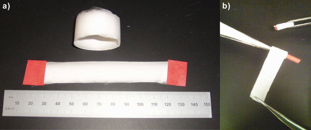
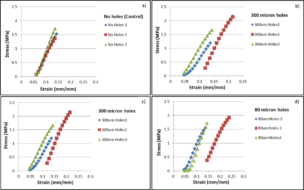
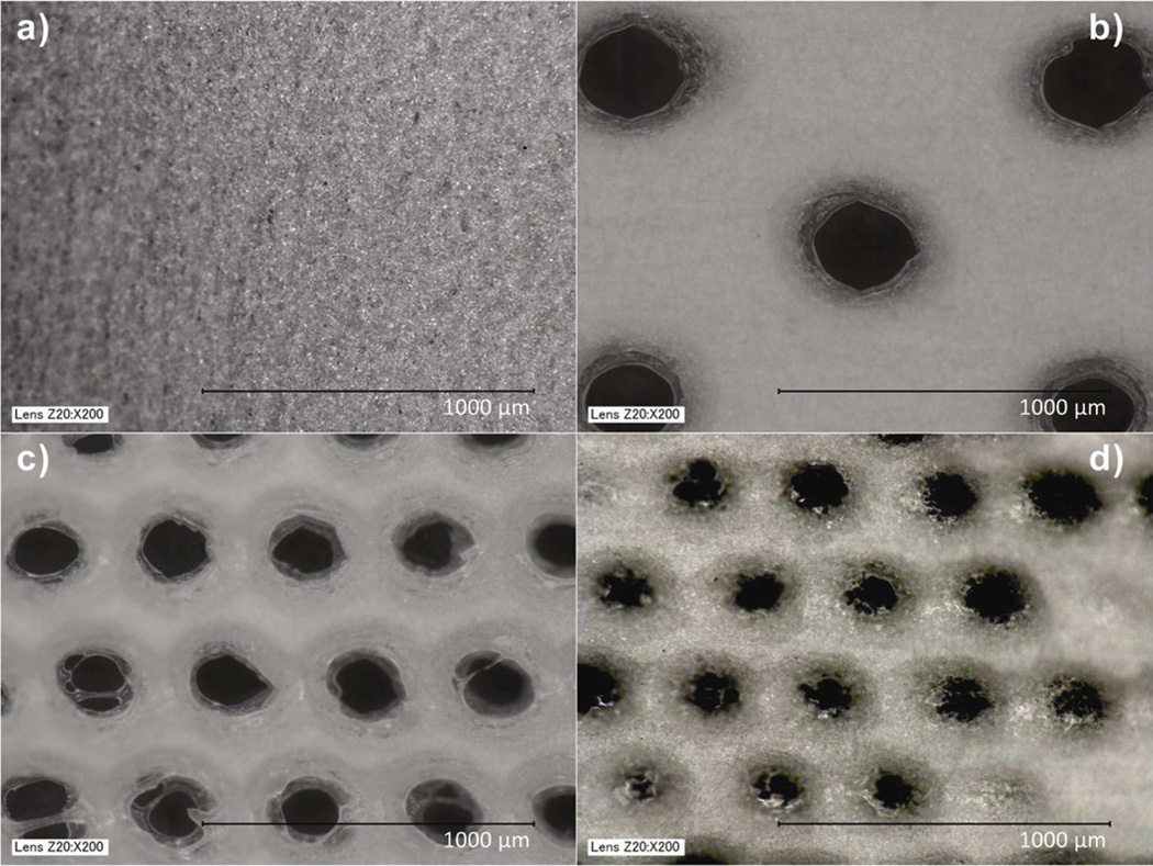
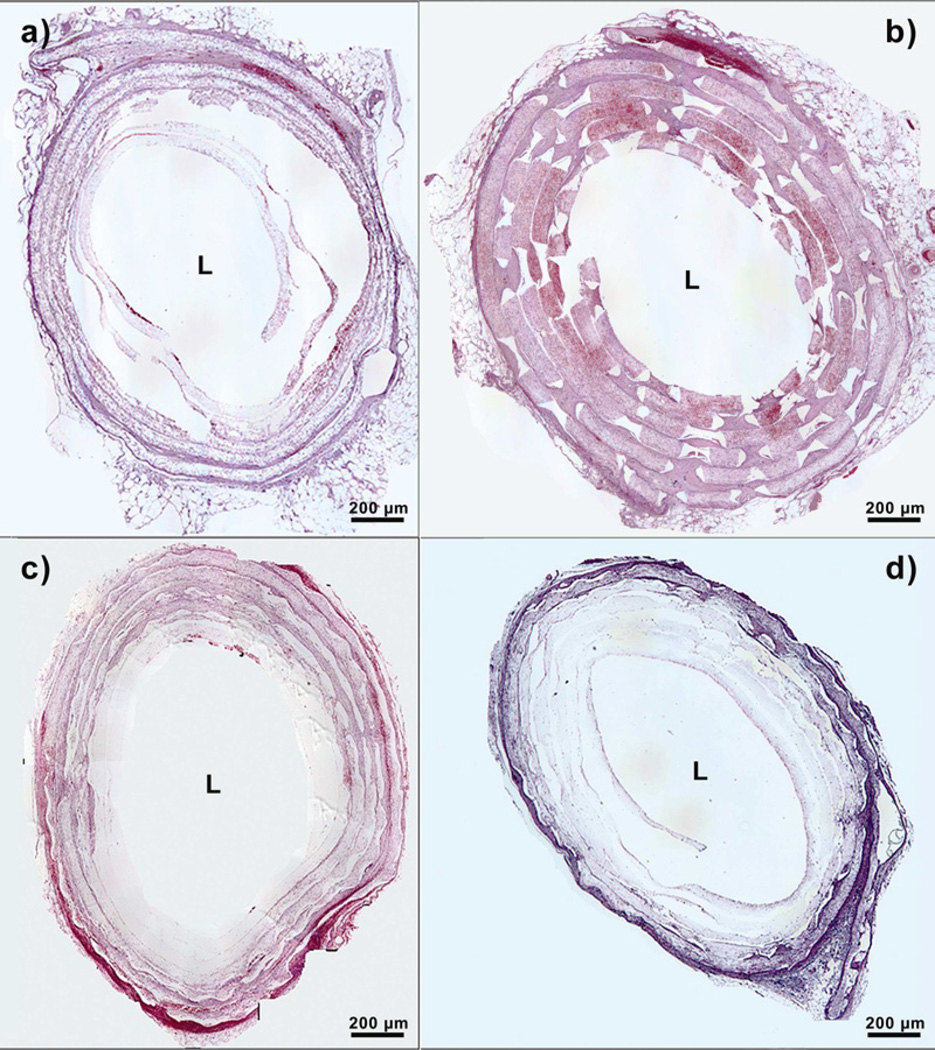
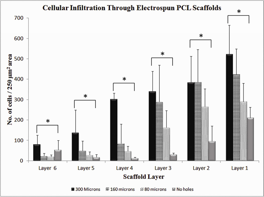
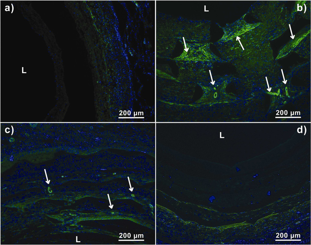
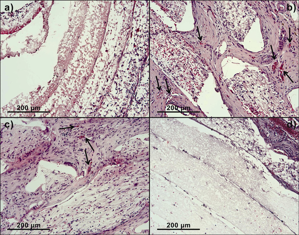
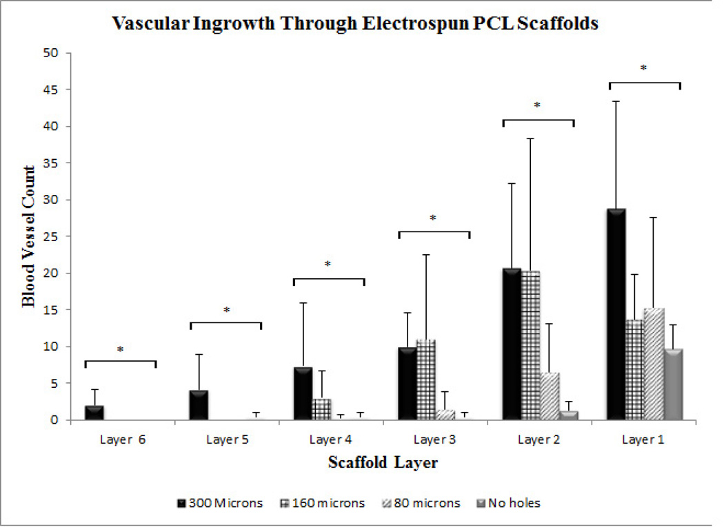
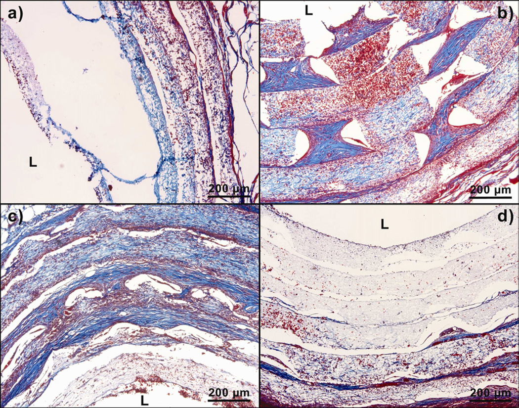
Polycaprolactone dissolved in hexafluoroisopropanol was electrospun at 25 kV to create uniform 100-120 μm sheets of polycaprolactone fiber mats (1- to 5-μm fiber diameter) with an array of pores created using VERSA LASER CUTTER 2.3. Three groups of fiber mats with three distinct pore diameters (300, 160, and 80 μm, all with 15% pore area) were fabricated and compared with a control group without laser cut pores. After laser cutting, all mats were collagen coated and manually wrapped around a catheter six times to form six concentric layers before implantation into the omentum of Lewis rats. Cellular infiltration and vascular ingrowth were examined after 2 wk.

RESULTS

Histologic analysis of 14-d samples showed that scaffolds with laser cut pores had close to 40% more cellular infiltration and increased vascular ingrowth in the innermost layers of the construct compared with the control group. Despite keeping pore area percentage constant between the three groups, the sheets with the largest pore size performed better than those with the smallest pore sizes.

CONCLUSIONS

Porosity is the primary factor limiting the extensive use of electrospun scaffolds in tissue engineering. Our method of LASER cutting pores in electrospun fibrous scaffolds ensures uniform pore sizes, easily controllable and customizable pores, and enhances cellular infiltration and vascular ingrowth, demonstrating significant advancement toward utility of electrospun scaffolds in tissue engineering.

摘要

背景

支架组织工程中最大的挑战之一仍然是细胞难以有效渗透到支架中,从而生成厚的血管化和细胞组织。静电纺丝已成为一种生产具有高表面积与体积比并类似于细胞外基质的支架的首选方法。然而,由于高纤维密度的静电纺丝支架缺乏大孔连通性,细胞渗透和血管生成仍然不足。在这项研究中,我们报告了一种新颖的两步静电纺丝和激光切割制造方法,以增强静电纺丝支架的大孔性。

材料与方法

将聚己内酯溶解在六氟异丙醇中,在 25 kV 下进行静电纺丝,以创建均匀的 100-120 μm 聚己内酯纤维垫(纤维直径为 1-5 μm),并使用 VERSA LASER CUTTER 2.3 制造出一系列孔阵列。制造了三组纤维垫,具有三种不同的孔径(300、160 和 80 μm,所有孔径的孔隙率均为 15%),并与未激光切割孔的对照组进行了比较。激光切割后,所有的垫子都用胶原蛋白涂层,并手动包裹在导管上六次,形成六个同心层,然后植入到 Lewis 大鼠的大网膜中。在植入后 2 周检查细胞渗透和血管生成。

结果

14 天样本的组织学分析表明,与对照组相比,激光切割孔的支架具有近 40%更多的细胞渗透,并且在构建物的最内层增加了血管生成。尽管三组之间保持相同的孔面积百分比,但具有最大孔径的薄片比具有最小孔径的薄片表现更好。

结论

孔隙率是限制静电纺丝支架在组织工程中广泛应用的主要因素。我们在静电纺丝纤维支架中激光切割孔的方法确保了均匀的孔径、易于控制和可定制的孔以及增强了细胞渗透和血管生成,这表明在组织工程中使用静电纺丝支架取得了重大进展。

https://cdn.ncbi.nlm.nih.gov/pmc/blobs/a2a0/3694756/4d6aa637b43f/nihms433663f9.jpg
https://cdn.ncbi.nlm.nih.gov/pmc/blobs/a2a0/3694756/daad4d4fd055/nihms433663f1.jpg
https://cdn.ncbi.nlm.nih.gov/pmc/blobs/a2a0/3694756/eea173cb4069/nihms433663f2.jpg
https://cdn.ncbi.nlm.nih.gov/pmc/blobs/a2a0/3694756/885ea8e4eba8/nihms433663f3.jpg
https://cdn.ncbi.nlm.nih.gov/pmc/blobs/a2a0/3694756/210273f1c401/nihms433663f4.jpg
https://cdn.ncbi.nlm.nih.gov/pmc/blobs/a2a0/3694756/4cb355f31d8b/nihms433663f5.jpg
https://cdn.ncbi.nlm.nih.gov/pmc/blobs/a2a0/3694756/1b7059faa969/nihms433663f6.jpg
https://cdn.ncbi.nlm.nih.gov/pmc/blobs/a2a0/3694756/543e64e81372/nihms433663f7.jpg
https://cdn.ncbi.nlm.nih.gov/pmc/blobs/a2a0/3694756/ec0bb38444de/nihms433663f8.jpg
https://cdn.ncbi.nlm.nih.gov/pmc/blobs/a2a0/3694756/4d6aa637b43f/nihms433663f9.jpg

相似文献

1
Macroporosity enhances vascularization of electrospun scaffolds.大孔结构增强静电纺丝支架的血管化。
J Surg Res. 2013 Jul;183(1):18-26. doi: 10.1016/j.jss.2013.01.005. Epub 2013 Feb 1.
2
Increasing the pore size of electrospun scaffolds.增加静电纺丝支架的孔径。
Tissue Eng Part B Rev. 2011 Oct;17(5):365-72. doi: 10.1089/ten.teb.2011.0235. Epub 2011 Aug 4.
3
Increasing the pore sizes of bone-mimetic electrospun scaffolds comprised of polycaprolactone, collagen I and hydroxyapatite to enhance cell infiltration.增大由聚己内酯、I 型胶原和羟基磷灰石组成的仿生电纺支架的孔径以增强细胞浸润。
Biomaterials. 2012 Jan;33(2):524-34. doi: 10.1016/j.biomaterials.2011.09.080. Epub 2011 Oct 19.
4
Cryogenic electrospinning: proposed mechanism, process parameters and its use in engineering of bilayered tissue structures.低温静电纺丝:提出的机制、工艺参数及其在双层组织工程中的应用。
Nanomedicine (Lond). 2013 Apr;8(4):555-66. doi: 10.2217/nnm.13.39.
5
Electrospun bilayer fibrous scaffolds for enhanced cell infiltration and vascularization in vivo.用于增强体内细胞浸润和血管生成的电纺双层纤维支架。
Acta Biomater. 2015 Feb;13:131-41. doi: 10.1016/j.actbio.2014.11.014. Epub 2014 Nov 15.
6
Gradient porous fibrous scaffolds: a novel approach to improving cell penetration in electrospun scaffolds.梯度多孔纤维支架:提高静电纺丝支架细胞渗透性的新方法。
Biomed Mater. 2018 Sep 21;13(6):065010. doi: 10.1088/1748-605X/aadbbe.
7
Fabrication of large pores in electrospun nanofibrous scaffolds for cellular infiltration: a review.静电纺丝纳米纤维支架中大孔的制备及其对细胞渗透的影响:综述。
Tissue Eng Part B Rev. 2012 Apr;18(2):77-87. doi: 10.1089/ten.TEB.2011.0390. Epub 2011 Dec 14.
8
Fabrication of three-dimensional porous scaffolds with controlled filament orientation and large pore size via an improved E-jetting technique.通过改进的电子喷射技术制备具有可控长丝取向和大孔径的三维多孔支架。
J Biomed Mater Res B Appl Biomater. 2014 May;102(4):651-8. doi: 10.1002/jbm.b.33043. Epub 2013 Oct 24.
9
Electrospun polycaprolactone scaffolds with tailored porosity using two approaches for enhanced cellular infiltration.采用两种方法构建具有定制孔隙率的静电纺聚己内酯支架,以增强细胞渗透。
J Mater Sci Mater Med. 2013 Jan;24(1):179-87. doi: 10.1007/s10856-012-4771-7. Epub 2012 Sep 29.
10
In vitro cell infiltration and in vivo cell infiltration and vascularization in a fibrous, highly porous poly(D,L-lactide) scaffold fabricated by cryogenic electrospinning technique.通过低温静电纺丝技术制备的纤维状、高度多孔聚(D,L-丙交酯)支架中的体外细胞浸润以及体内细胞浸润和血管生成。
J Biomed Mater Res A. 2009 Oct;91(1):231-40. doi: 10.1002/jbm.a.32208.

引用本文的文献

1
Preclinical Evaluation and Advancements in Vascularized Bone Tissue Engineering.血管化骨组织工程的临床前评估与进展
Biomimetics (Basel). 2025 Jun 20;10(7):412. doi: 10.3390/biomimetics10070412.
2
Biofabrication of engineered blood vessels for biomedical applications.用于生物医学应用的工程血管的生物制造。
Sci Technol Adv Mater. 2024 Mar 21;25(1):2330339. doi: 10.1080/14686996.2024.2330339. eCollection 2024.
3
Risedronate-loaded aerogel scaffolds for bone regeneration.载雷奈酸锶的气凝胶支架用于骨再生。

本文引用的文献

1
Lamellar stack formation and degradative behaviors of hydrolytically degraded poly(ε-caprolactone) and poly(glycolide-ε-caprolactone) blended fibers.层状堆积形成和水解降解聚(ε-己内酯)和聚(乙交酯-ε-己内酯)共混纤维的降解行为。
J Biomed Mater Res B Appl Biomater. 2012 Jan;100(1):274-84. doi: 10.1002/jbm.b.31950. Epub 2011 Nov 9.
2
Tuning electrospinning parameters for production of 3D-fiber-fleeces with increased porosity for soft tissue engineering applications.调整静电纺丝参数,以生产具有更高孔隙率的 3D 纤维绒毛,用于软组织工程应用。
Eur Cell Mater. 2011 Mar 22;21:286-303. doi: 10.22203/ecm.v021a22.
3
Increasing electrospun scaffold pore size with tailored collectors for improved cell penetration.
Drug Deliv. 2023 Dec;30(1):51-63. doi: 10.1080/10717544.2022.2152135.
4
Growth Factor-Free Vascularization of Marine-Origin Collagen Sponges Using Cryopreserved Stromal Vascular Fractions from Human Adipose Tissue.利用人脂肪组织冷冻保存的基质血管成分实现海洋来源胶原海绵的无生长因子血管化。
Mar Drugs. 2022 Sep 30;20(10):623. doi: 10.3390/md20100623.
5
Prospects and Challenges of Electrospun Cell and Drug Delivery Vehicles to Correct Urethral Stricture.电纺细胞和药物输送载体在纠正尿道狭窄中的前景和挑战。
Int J Mol Sci. 2022 Sep 10;23(18):10519. doi: 10.3390/ijms231810519.
6
In Vitro Strategies to Vascularize 3D Physiologically Relevant Models.体外策略使 3D 生理相关模型血管化。
Adv Sci (Weinh). 2021 Oct;8(19):e2100798. doi: 10.1002/advs.202100798. Epub 2021 Aug 5.
7
Angiogenic biomaterials to promote therapeutic regeneration and investigate disease progression.用于促进治疗性再生和研究疾病进展的血管生成生物材料。
Biomaterials. 2020 Oct;255:120207. doi: 10.1016/j.biomaterials.2020.120207. Epub 2020 Jun 14.
8
Solution Blow Spinning of Polyvinylidene Fluoride Based Fibers for Energy Harvesting Applications: A Review.用于能量收集应用的聚偏氟乙烯基纤维的溶液吹纺:综述
Polymers (Basel). 2020 Jun 7;12(6):1304. doi: 10.3390/polym12061304.
9
Random auxetics from buckling fibre networks.随机屈曲纤维网络的各向异性材料。
Nat Commun. 2019 Oct 25;10(1):4863. doi: 10.1038/s41467-019-12757-7.
10
Fabrication and Characterisation of Stimuli Responsive Piezoelectric PVDF and Hydroxyapatite-Filled PVDF Fibrous Membranes.刺激响应型压电 PVDF 及羟基磷灰石填充 PVDF 纤维膜的制备与表征。
通过定制收集器增加静电纺丝支架的孔径,以提高细胞穿透性。
Acta Biomater. 2011 Jun;7(6):2544-57. doi: 10.1016/j.actbio.2011.02.036. Epub 2011 Mar 1.
4
Controlling the porosity of fibrous scaffolds by modulating the fiber diameter and packing density.通过调节纤维直径和堆积密度来控制纤维支架的孔隙率。
J Biomed Mater Res A. 2011 Mar 1;96(3):566-74. doi: 10.1002/jbm.a.33010. Epub 2011 Jan 10.
5
Pre-osteoblast infiltration and differentiation in highly porous apatite-coated PLLA electrospun scaffolds.高度多孔磷灰石涂层 PLLA 电纺支架中的成前体细胞的浸润和分化。
Biomaterials. 2011 Mar;32(9):2294-304. doi: 10.1016/j.biomaterials.2010.12.003. Epub 2010 Dec 30.
6
Effect of fiber diameter, pore size and seeding method on growth of human dermal fibroblasts in electrospun poly(epsilon-caprolactone) fibrous mats.纤维直径、孔径和接种方法对聚己内酯纤维垫中人真皮成纤维细胞生长的影响。
Biomaterials. 2010 Jan;31(3):491-504. doi: 10.1016/j.biomaterials.2009.09.072. Epub 2009 Oct 12.
7
Improved infiltration of stem cells on electrospun nanofibers.干细胞在电纺纳米纤维上的浸润性得到改善。
Biochem Biophys Res Commun. 2009 Apr 24;382(1):129-33. doi: 10.1016/j.bbrc.2009.02.150. Epub 2009 Mar 3.
8
In vitro cell infiltration and in vivo cell infiltration and vascularization in a fibrous, highly porous poly(D,L-lactide) scaffold fabricated by cryogenic electrospinning technique.通过低温静电纺丝技术制备的纤维状、高度多孔聚(D,L-丙交酯)支架中的体外细胞浸润以及体内细胞浸润和血管生成。
J Biomed Mater Res A. 2009 Oct;91(1):231-40. doi: 10.1002/jbm.a.32208.
9
Electrospun nanofiber scaffolds: engineering soft tissues.电纺纳米纤维支架:软组织工程
Biomed Mater. 2008 Sep;3(3):034002. doi: 10.1088/1748-6041/3/3/034002. Epub 2008 Aug 8.
10
Electrospun fibrous mats with high porosity as potential scaffolds for skin tissue engineering.具有高孔隙率的电纺纤维垫作为皮肤组织工程的潜在支架。
Biomacromolecules. 2008 Jul;9(7):1795-801. doi: 10.1021/bm800476u. Epub 2008 Jun 26.