Brain Research Imaging Centre (BRIC), Neuroimaging Sciences, University of Edinburgh, Western General Hospital, Crewe Road, Edinburgh EH4 2XU, UK.
Neuroimage. 2013 Nov 15;82:470-80. doi: 10.1016/j.neuroimage.2013.06.013. Epub 2013 Jun 12.
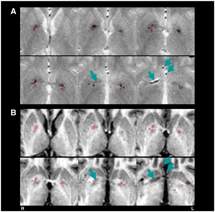
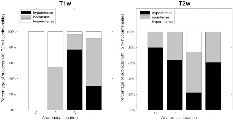
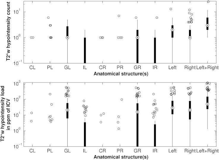
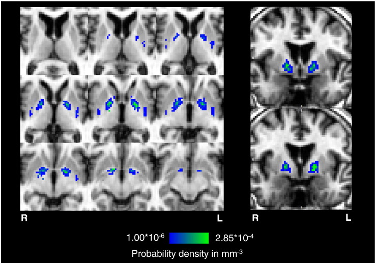
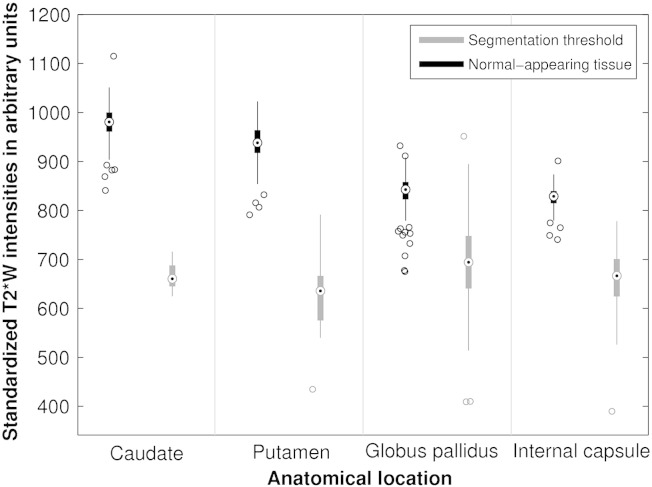
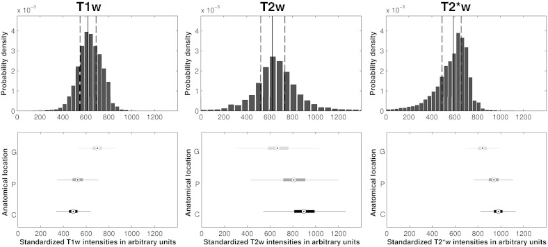
Multifocal T2*-weighted (T2w) hypointensities in the basal ganglia, which are believed to arise predominantly from mineralized small vessels and perivascular spaces, have been proposed as a biomarker for cerebral small vessel disease. This study provides baseline data on their appearance on conventional structural MRI for improving and automating current manual segmentation methods. Using a published thresholding method, multifocal T2w hypointensities were manually segmented from whole brain T2w volumes acquired from 98 community-dwelling subjects in their early 70s. Connected component analysis was used to derive the average T2w hypointensity count and load per basal ganglia nucleus, as well as the morphology of their connected components, while nonlinear spatial probability mapping yielded their spatial distribution. T1-weighted (T1w), T2-weighted (T2w) and T2w intensity distributions of basal ganglia T2w hypointensities and their appearance on T1w and T2w MRI were investigated to gain further insights into the underlying tissue composition. In 75/98 subjects, on average, 3 T2w hypointensities with a median total volume per intracranial volume of 50.3ppm were located in and around the globus pallidus. Individual hypointensities appeared smooth and spherical with a median volume of 12mm(3) and median in-plane area of 4mm(2). Spatial probability maps suggested an association between T2w hypointensities and the point of entry of lenticulostriate arterioles into the brain parenchyma. T1w and T2w and especially the T2w intensity distributions of these hypointensities, which were negatively skewed, were generally not normally distributed indicating an underlying inhomogeneous tissue structure. Globus pallidus T2w hypointensities tended to appear hypo- and isointense on T1w and T2w MRI, whereas those from other structures appeared iso- and hypointense. This pattern could be explained by an increased mineralization of the globus pallidus. In conclusion, the characteristic spatial distribution and appearance of multifocal basal ganglia T2*w hypointensities in our elderly cohort on structural MRI appear to support the suggested association with mineralized proximal lenticulostriate arterioles and perivascular spaces.
基底节区多灶性 T2*-加权(T2w)低信号,据信主要来自矿化小血管和血管周围间隙,已被提议作为脑小血管疾病的生物标志物。本研究为改善和自动化当前的手动分割方法,提供了其在常规结构 MRI 上的出现的基线数据。使用已发表的阈值方法,从 98 名 70 岁出头的社区居住者的全脑 T2w 容积中手动分割多灶性 T2w 低信号。连通分量分析用于得出每个基底节核团的 T2w 低信号平均计数和负荷,以及它们连通分量的形态,而非线性空间概率映射则产生它们的空间分布。研究了基底节区 T2w 低信号的 T1 加权(T1w)、T2 加权(T2w)和 T2w 强度分布及其在 T1w 和 T2w MRI 上的表现,以进一步深入了解潜在的组织成分。在 98 名受试者中的 75 名中,平均有 3 个 T2w 低信号,每个颅内容积的中位数总容积为 50.3ppm,位于苍白球内和周围。单个低信号表现为光滑和球形,中位数体积为 12mm(3),中位数平面面积为 4mm(2)。空间概率图表明 T2w 低信号与纹状体动脉进入脑实质的进入点之间存在关联。T1w 和 T2w,特别是这些低信号的 T2w 强度分布,呈负偏态,通常不符合正态分布,表明存在潜在的不均匀组织结构。苍白球 T2w 低信号在 T1w 和 T2w MRI 上倾向于表现为低信号和等信号,而其他结构的低信号则表现为等信号和低信号。这种模式可以通过苍白球的矿化增加来解释。总之,我们老年队列在结构 MRI 上多灶性基底节区 T2*w 低信号的特征性空间分布和表现似乎支持与矿化近端纹状体动脉和血管周围间隙的建议关联。