Sezione di Anatomia Umana, Dipartimento di Biomedicina Sperimentale e Neuroscienze Cliniche, Università di Palermo, Palermo, Italia.
Eur J Histochem. 2013 Jun 28;57(2):e20. doi: 10.4081/ejh.2013.e20.
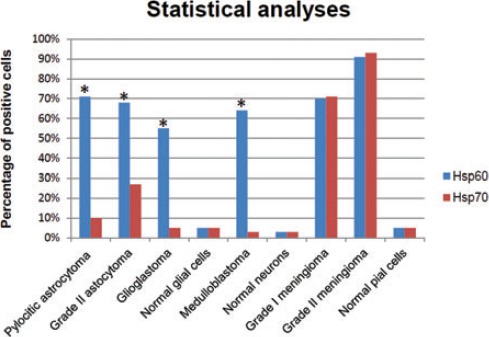
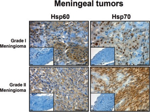
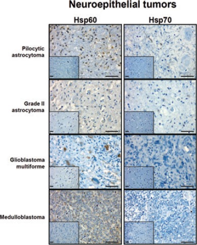
In this work we analysed, by immunohistochemistry, a series of brain tumors to detect the levels and cellular distribution of Hsp60 and Hsp70. We found that Hsp60 levels were significantly higher than those of Hsp70 in neuroepithelial tumors, while levels of both molecules were not significantly different from each other in meningeal neoplasms. In particular, Hsp60 immunopositivity was present mainly at the cytoplasmic level, while Hsp70 immunopositivity was found both in the cytoplasm and in the nucleus of tumor cells. The levels of these molecules in healthy control cells were always very low. Finally, Hsp60 and Hsp70 levels did not correlate with the different types (WHO grade) of neoplasm. Our results are partially in agreement with previous studies and suggest that Hsp60 is not increased by a passive phenomenon (e.g., due to the stress caused by the peritumor environment on cancer cells) but may be actively implicated in tumor progression, e.g. inhibiting tumor cell death or antitumor immune system response, as already postulated in vitro. We also briefly discuss the most recent publications on the extramitochondrial localization of Hsp60 in tumor cells and its role in tumor progression.
在这项工作中,我们通过免疫组织化学分析了一系列脑肿瘤,以检测 Hsp60 和 Hsp70 的水平和细胞分布。我们发现,神经上皮肿瘤中 Hsp60 的水平明显高于 Hsp70,而脑膜肿瘤中两种分子的水平彼此之间没有显著差异。特别是,Hsp60 免疫阳性主要位于细胞质水平,而 Hsp70 免疫阳性则存在于肿瘤细胞的细胞质和细胞核中。这些分子在健康对照细胞中的水平始终非常低。最后,这些分子的水平与不同类型(WHO 分级)的肿瘤无关。我们的结果与之前的一些研究部分一致,表明 Hsp60 的增加不是被动现象(例如,由于肿瘤周围环境对癌细胞造成的应激)引起的,而是可能主动参与肿瘤的进展,例如抑制肿瘤细胞死亡或抗肿瘤免疫系统反应,正如已经在体外提出的那样。我们还简要讨论了最近关于 Hsp60 在肿瘤细胞中线粒体外定位及其在肿瘤进展中的作用的出版物。