Zhong Qisheng, Yuan Shaoji
Department of Neurosurgery, General Hospital of Jinan Military Command of Chinese PLA, Jinan, Shandong 250031, P.R. China.
Oncol Lett. 2013 Jun;5(6):1783-1786. doi: 10.3892/ol.2013.1293. Epub 2013 Apr 8.
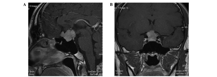
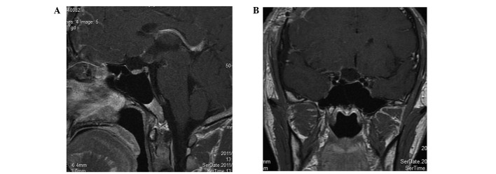
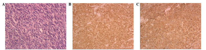
The present study reports the case of a patient with a vision impairment in the right eye. Head computed tomography revealed a round, hyperdense mass in the sellar and suprasellar regions. Pituitary gland magnetic resonance imaging (MRI) revealed isointensity on T1- and T2-weighted imaging. Tumor-enhanced scanning showed heterogeneous contrast enhancement. The initial diagnosis was that of meningioma or pituitary tumor. A total tumor resection was performed using a right pterional approach under general anesthesia. During surgery, the base of the tumor was located on the sellar diaphragm of the left anterior pituitary stalk. The pathological diagnosis was of a solitary fibrous tumor (SFT). The patient had no post-operative diabetes insipidus or idiopathic pituitary hypofunction. The clinical experience, imaging information and pathological features of SFT in this case report may provide a reference for correct diagnosis and total resection of SFTs in the sella turcica.
本研究报告了一例右眼视力受损的患者。头部计算机断层扫描显示蝶鞍区和鞍上区有一个圆形的高密度肿块。垂体磁共振成像(MRI)在T1加权和T2加权成像上显示等信号。肿瘤增强扫描显示不均匀强化。初步诊断为脑膜瘤或垂体瘤。在全身麻醉下采用右侧翼点入路进行肿瘤全切。手术中,肿瘤基底位于左前垂体柄的鞍膈上。病理诊断为孤立性纤维瘤(SFT)。患者术后无尿崩症或特发性垂体功能减退。本病例报告中SFT的临床经验、影像学信息和病理特征可为蝶鞍区SFT的正确诊断和全切提供参考。