• 文献检索
  • 文档翻译
  • 深度研究
  • 学术资讯
  • Suppr Zotero 插件Zotero 插件
  • 邀请有礼
  • 套餐&价格
  • 历史记录
应用&插件
Suppr Zotero 插件Zotero 插件浏览器插件Mac 客户端Windows 客户端微信小程序
定价
高级版会员购买积分包购买API积分包
服务
文献检索文档翻译深度研究API 文档MCP 服务
关于我们
关于 Suppr公司介绍联系我们用户协议隐私条款
关注我们

Suppr 超能文献

核心技术专利:CN118964589B侵权必究
粤ICP备2023148730 号-1Suppr @ 2026

文献检索

告别复杂PubMed语法,用中文像聊天一样搜索,搜遍4000万医学文献。AI智能推荐,让科研检索更轻松。

立即免费搜索

文件翻译

保留排版,准确专业,支持PDF/Word/PPT等文件格式,支持 12+语言互译。

免费翻译文档

深度研究

AI帮你快速写综述,25分钟生成高质量综述,智能提取关键信息,辅助科研写作。

立即免费体验

吸气期CT扫描正常的患者进行呼气期CT扫描:闭塞性细支气管炎及其他细支气管阻塞原因的一项发现。

Expiratory CT scan in patients with normal inspiratory CT scan: a finding of obliterative bronchiolitis and other causes of bronchiolar obstruction.

作者信息

Gaeta Michele, Minutoli Fabio, Girbino Giuseppe, Murabito Alessandra, Benedetto Caterina, Contiguglia Rosario, Ruggeri Paolo, Privitera Salvatore

机构信息

Department of Biomedical Sciences and of Morphological and Functional Images, University of Messina, Messina, Italy.

出版信息

Multidiscip Respir Med. 2013 Jul 9;8(1):44. doi: 10.1186/2049-6958-8-44.

DOI:10.1186/2049-6958-8-44
PMID:23835554
原文链接:https://pmc.ncbi.nlm.nih.gov/articles/PMC3710098/
Abstract

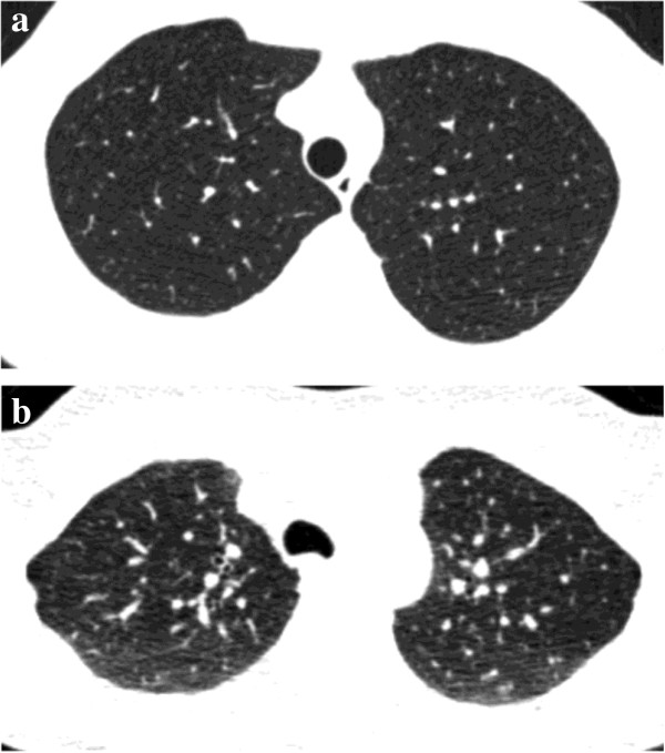
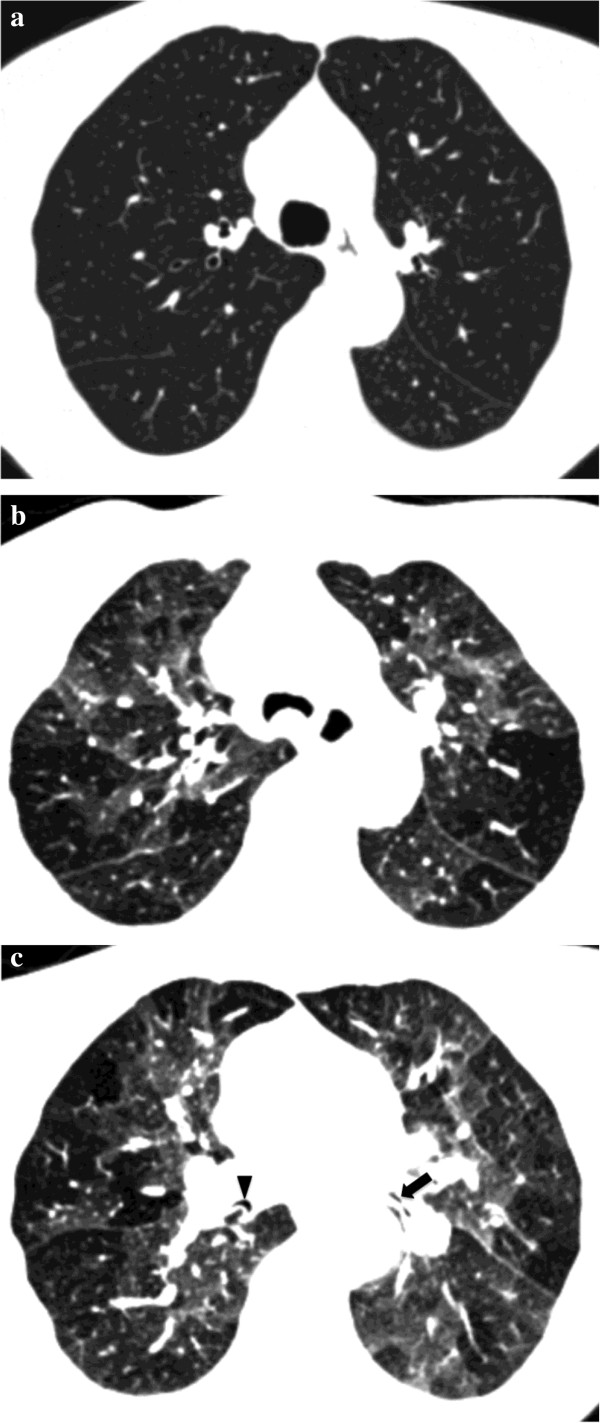
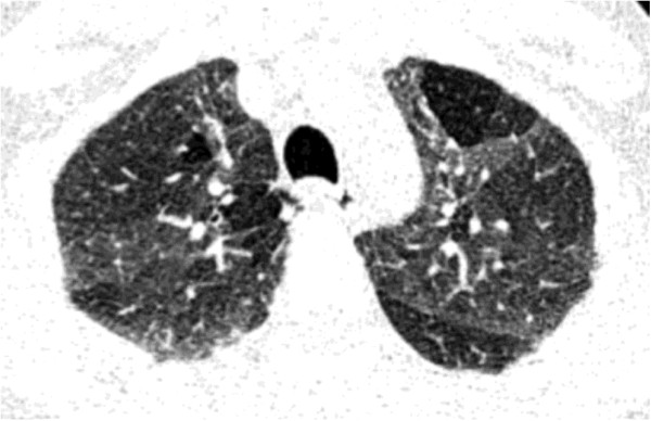
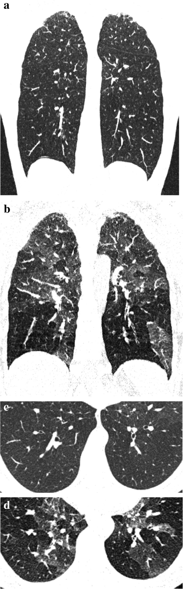
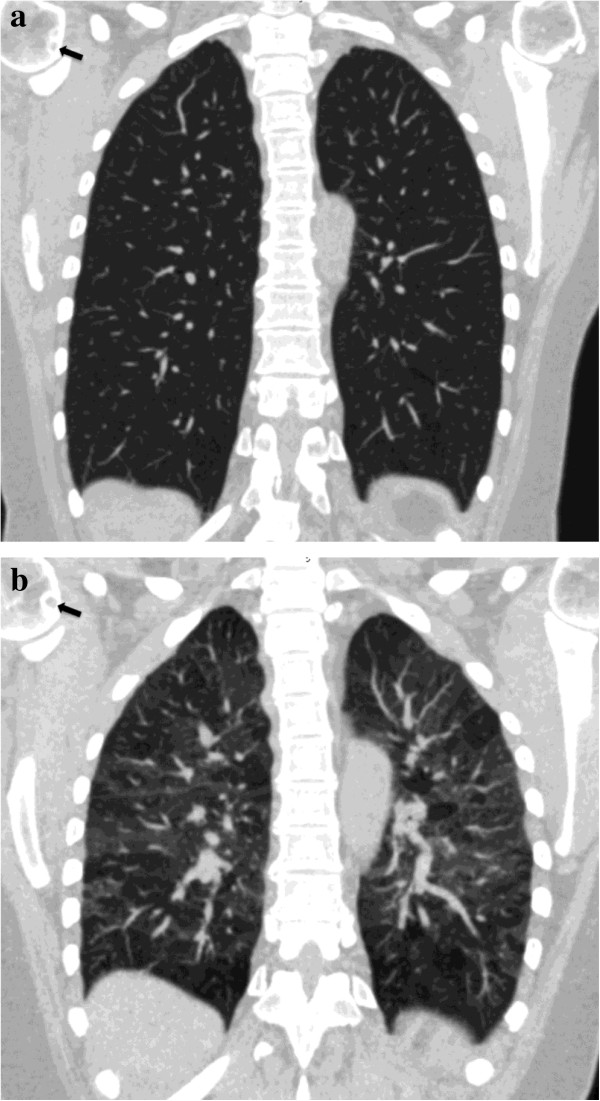
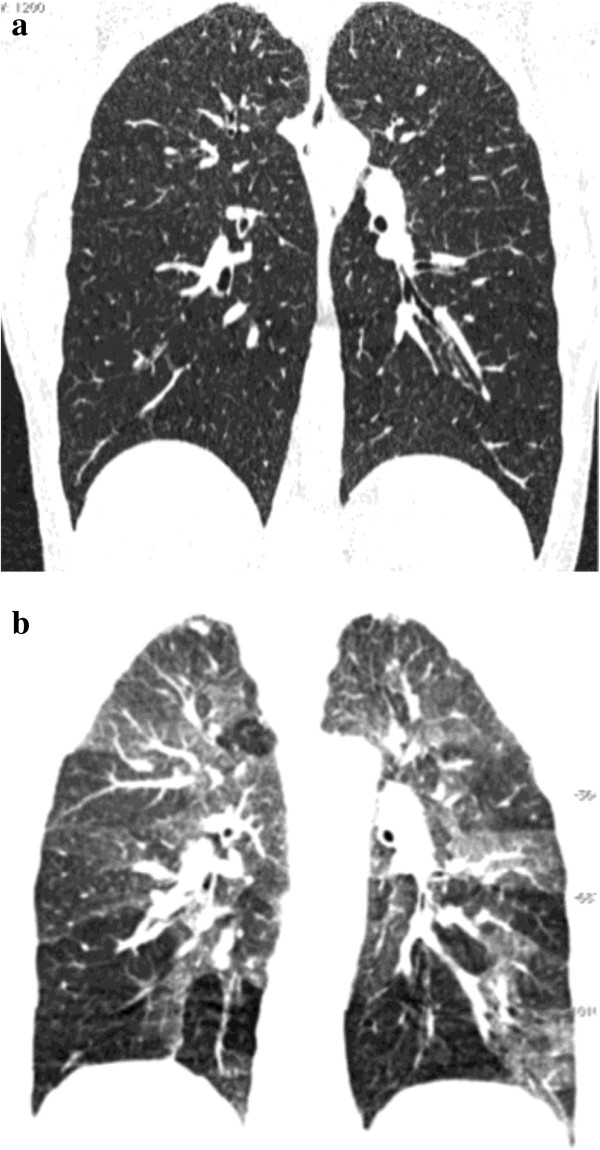
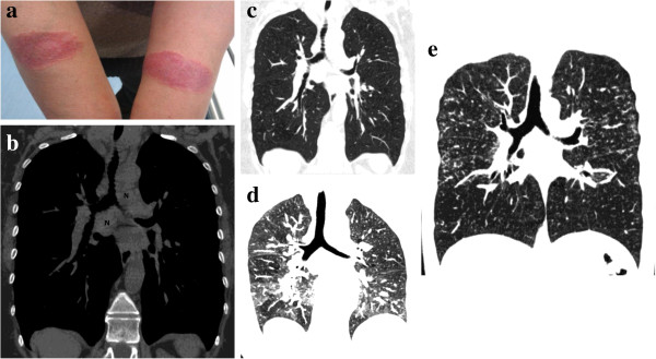
Expiratory CT scan is usually obtained as supplement to normal inspiratory CT scan to recognize air-trapping, which is expression of small airways obstruction. In some patients the air-trapping may be the only sign of an early-stage small airways disease in an otherwise normal lung.The purpose of this article is to illustrate pathologic conditions, namely obliterative bronchiolitis, in which expiratory CT scan can be abnormal despite normal inspiratory CT examination, and to highlight indications for this technique in patients with clinical and functional suspect of bronchiolar obstruction.

摘要

呼气期CT扫描通常作为常规吸气期CT扫描的补充,用于识别气体陷闭,这是小气道阻塞的表现。在一些患者中,气体陷闭可能是原本正常的肺中早期小气道疾病的唯一征象。本文旨在阐述一些病理情况,即闭塞性细支气管炎,在这些病例中,尽管吸气期CT检查正常,但呼气期CT扫描仍可能出现异常,并强调该技术在临床上怀疑有细支气管阻塞且有功能异常的患者中的应用指征。

https://cdn.ncbi.nlm.nih.gov/pmc/blobs/7f36/3710098/a72b7fbdf7ff/2049-6958-8-44-8.jpg
https://cdn.ncbi.nlm.nih.gov/pmc/blobs/7f36/3710098/77151ce5e992/2049-6958-8-44-1.jpg
https://cdn.ncbi.nlm.nih.gov/pmc/blobs/7f36/3710098/3d7af1f86e17/2049-6958-8-44-2.jpg
https://cdn.ncbi.nlm.nih.gov/pmc/blobs/7f36/3710098/09f9a6a6e37d/2049-6958-8-44-3.jpg
https://cdn.ncbi.nlm.nih.gov/pmc/blobs/7f36/3710098/c0a7a0617397/2049-6958-8-44-4.jpg
https://cdn.ncbi.nlm.nih.gov/pmc/blobs/7f36/3710098/441bfb300710/2049-6958-8-44-5.jpg
https://cdn.ncbi.nlm.nih.gov/pmc/blobs/7f36/3710098/e3a793dbd9f9/2049-6958-8-44-6.jpg
https://cdn.ncbi.nlm.nih.gov/pmc/blobs/7f36/3710098/2ca1c952cc8b/2049-6958-8-44-7.jpg
https://cdn.ncbi.nlm.nih.gov/pmc/blobs/7f36/3710098/a72b7fbdf7ff/2049-6958-8-44-8.jpg

相似文献

1
Expiratory CT scan in patients with normal inspiratory CT scan: a finding of obliterative bronchiolitis and other causes of bronchiolar obstruction.吸气期CT扫描正常的患者进行呼气期CT扫描:闭塞性细支气管炎及其他细支气管阻塞原因的一项发现。
Multidiscip Respir Med. 2013 Jul 9;8(1):44. doi: 10.1186/2049-6958-8-44.
2
[Dynamic computed tomography in the study of bronchiolitis obliterans].[动态计算机断层扫描在闭塞性细支气管炎研究中的应用]
Radiol Med. 1997 Oct;94(4):308-14.
3
Air trapping on expiratory high-resolution CT scans in the absence of inspiratory scan abnormalities: correlation with pulmonary function tests and differential diagnosis.呼气期高分辨率CT扫描显示空气潴留而吸气期扫描无异常:与肺功能检查的相关性及鉴别诊断
AJR Am J Roentgenol. 1998 May;170(5):1349-53. doi: 10.2214/ajr.170.5.9574614.
4
Obliterative bronchiolitis: individual CT signs of small airways disease and functional correlation.闭塞性细支气管炎:小气道疾病的个体CT征象及功能相关性
Radiology. 1997 Jun;203(3):721-6. doi: 10.1148/radiology.203.3.9169694.
5
Obstructive flow-volume loop contours after single lung transplantation.单肺移植后的阻塞性流量-容积环轮廓
J Heart Lung Transplant. 1994 May-Jun;13(3):508-13.
6
Expiratory CT scan: When to do it and how to interpret it.呼气期CT扫描:何时进行及如何解读
Radiologia (Engl Ed). 2023 Jul-Aug;65(4):352-361. doi: 10.1016/j.rxeng.2023.01.008. Epub 2023 Jul 12.
7
Air trapping in primary Sjögren syndrome: correlation of expiratory CT with pulmonary function tests.原发性干燥综合征中的空气潴留:呼气CT与肺功能测试的相关性。
J Comput Assist Tomogr. 1999 Mar-Apr;23(2):169-73. doi: 10.1097/00004728-199903000-00002.
8
Correlation of high-resolution CT and pulmonary function in bronchiolitis obliterans: a study based on 24 patients associated with consumption of Sauropus androgynus.闭塞性细支气管炎的高分辨率CT与肺功能的相关性:一项基于24例与食用雷公藤相关患者的研究。
AJR Am J Roentgenol. 1997 Apr;168(4):1045-50. doi: 10.2214/ajr.168.4.9124113.
9
Post-lung transplantation bronchiolitis obliterans syndrome: usefulness of expiratory thin-section CT for diagnosis.肺移植术后闭塞性细支气管炎综合征:呼气期薄层CT在诊断中的应用价值
Radiology. 2001 Aug;220(2):455-62. doi: 10.1148/radiology.220.2.r01au19455.
10
Overview and Challenges of Bronchiolar Disorders.细支气管疾病概述及挑战。
Ann Am Thorac Soc. 2020 Mar;17(3):253-263. doi: 10.1513/AnnalsATS.201907-569CME.

引用本文的文献

1
Evaluating the Cumulative Benefit of Inspiratory CT, Expiratory CT, and Clinical Data for COPD Diagnosis and Staging through Deep Learning.通过深度学习评估吸气CT、呼气CT和临床数据对慢性阻塞性肺疾病诊断和分期的累积益处。
Radiol Cardiothorac Imaging. 2024 Dec;6(6):e240005. doi: 10.1148/ryct.240005.
2
ESR Essentials: imaging in fibrotic lung diseases-practice recommendations by the European Society of Thoracic Imaging.红细胞沉降率要点:纤维化性肺病的影像学——欧洲胸部影像学会的实践建议
Eur Radiol. 2025 Apr;35(4):2245-2255. doi: 10.1007/s00330-024-11054-2. Epub 2024 Sep 7.
3
Expiratory Venous Volume and Arterial Tortuosity are Associated with Disease Severity and Mortality Risk in Patients with COPD: Results from COSYCONET.

本文引用的文献

1
Assessment of lung volume collapsibility in chronic obstructive lung disease patients using CT.使用 CT 评估慢性阻塞性肺疾病患者的肺容积塌陷性。
Eur Radiol. 2013 Jun;23(6):1564-72. doi: 10.1007/s00330-012-2746-1. Epub 2013 Mar 15.
2
Early identification of small airways disease on lung cancer screening CT: comparison of current air trapping measures.肺癌筛查 CT 中小气道疾病的早期识别:当前空气潴留测量方法的比较。
Lung. 2012 Dec;190(6):629-33. doi: 10.1007/s00408-012-9422-8. Epub 2012 Oct 12.
3
Respiratory involvement in inflammatory bowel diseases.
COSYCONET 研究:COPD 患者的呼气末静脉容积和动脉迂曲与疾病严重程度和死亡风险相关。
Int J Chron Obstruct Pulmon Dis. 2024 Jul 1;19:1515-1529. doi: 10.2147/COPD.S458905. eCollection 2024.
4
Direct estimation of regional lung volume change from paired and single CT images using residual regression neural network.使用残差回归神经网络从配对和单张 CT 图像直接估计区域肺容积变化。
Med Phys. 2023 Sep;50(9):5698-5714. doi: 10.1002/mp.16365. Epub 2023 Mar 26.
5
MR imaging of the airways.气道的磁共振成像。
Br J Radiol. 2023 Jun 1;96(1146):20220630. doi: 10.1259/bjr.20220630. Epub 2023 Mar 3.
6
Ventilator-Assisted Inspiratory and Expiratory Breath-Hold Thoracic Computed Tomographic Scans Can Detect Dynamic and Static Airway Collapse in Dogs with Limited Agreement with Tracheobronchoscopy.呼吸机辅助吸气和呼气屏气胸部计算机断层扫描可检测犬的动态和静态气道塌陷,与气管支气管镜检查的一致性有限。
Animals (Basel). 2022 Nov 10;12(22):3091. doi: 10.3390/ani12223091.
7
Interstitial Lung Abnormalities at CT: Subtypes, Clinical Significance, and Associations with Lung Cancer.CT 下的肺间质异常:亚型、临床意义及与肺癌的相关性。
Radiographics. 2022 Nov-Dec;42(7):1925-1939. doi: 10.1148/rg.220073. Epub 2022 Sep 9.
8
Comparison of magnetic resonance imaging and computed tomography to measure preoperative parameters of children with pectus excavatum.磁共振成像与计算机断层扫描在测量漏斗胸患儿术前参数方面的比较。
Pediatr Investig. 2019 Jun 25;3(2):102-109. doi: 10.1002/ped4.12132. eCollection 2019 Jun.
9
High throughput image labeling on chest computed tomography by deep learning.通过深度学习实现胸部计算机断层扫描的高通量图像标注
J Med Imaging (Bellingham). 2020 Mar;7(2):024501. doi: 10.1117/1.JMI.7.2.024501. Epub 2020 Mar 20.
10
Serial peak flow measurements in allergic alveolitis.过敏性肺泡炎的连续呼气峰流量测量
Occup Med (Lond). 2015 Aug;65(6):505-6. doi: 10.1093/occmed/kqv099.
炎症性肠病中的呼吸系统受累情况。
Multidiscip Respir Med. 2010 Jun 30;5(3):173-82. doi: 10.1186/2049-6958-5-3-173.
4
MRI differentiation of pneumonia-like mucinous adenocarcinoma and infectious pneumonia.MRI 鉴别肺炎型黏液腺癌与感染性肺炎。
Eur J Radiol. 2012 Nov;81(11):3587-91. doi: 10.1016/j.ejrad.2011.12.022. Epub 2012 Jan 13.
5
Identification of chronic obstructive pulmonary disease in lung cancer screening computed tomographic scans.肺癌筛查 CT 扫描中慢性阻塞性肺疾病的识别。
JAMA. 2011 Oct 26;306(16):1775-81. doi: 10.1001/jama.2011.1531.
6
Assessing and treating small airways disease in asthma and chronic obstructive pulmonary disease.评估和治疗哮喘和慢性阻塞性肺疾病中的小气道疾病。
Ann Med. 2012 Mar;44(2):146-56. doi: 10.3109/07853890.2011.585656. Epub 2011 Jun 17.
7
CT of diffuse tracheal diseases.弥漫性气管疾病的CT检查
AJR Am J Roentgenol. 2011 Mar;196(3):W240-6. doi: 10.2214/AJR.09.4146.
8
Imaging of lung function using hyperpolarized helium-3 magnetic resonance imaging: Review of current and emerging translational methods and applications.使用超极化氦-3 磁共振成像进行肺功能成像:当前和新兴转化方法及应用的综述。
J Magn Reson Imaging. 2010 Dec;32(6):1398-408. doi: 10.1002/jmri.22375.
9
Pulmonary sarcoidosis: the 'Great Pretender'.肺结节病:“伪装大师”。
Clin Radiol. 2010 Aug;65(8):642-50. doi: 10.1016/j.crad.2010.03.004. Epub 2010 May 13.
10
Bronchiolar disorders: a clinical-radiological diagnostic algorithm.细支气管疾病:一种临床-放射学诊断算法。
Chest. 2010 Apr;137(4):938-51. doi: 10.1378/chest.09-0800.