School of Applied and Engineering Physics, Cornell University, Ithaca, New York 14853, USA.
J Biophotonics. 2013 Oct;6(10):815-20. doi: 10.1002/jbio.201300005. Epub 2013 Jul 10.
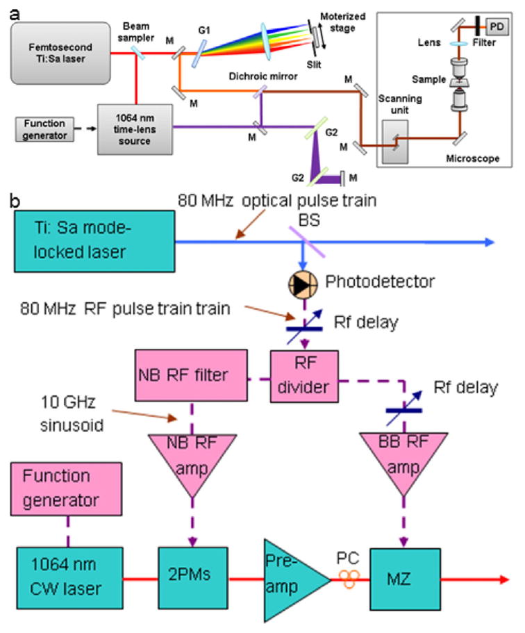
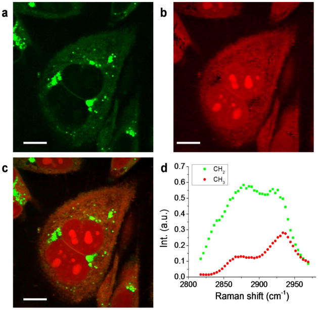
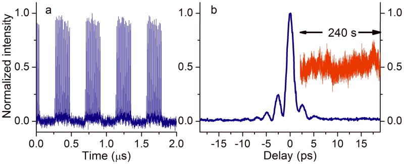
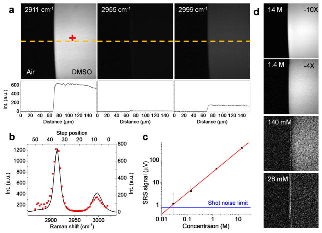
We demonstrate a hyperspectral stimulated Raman scattering (SRS) microscope through spectral-transformed excitation. The 1064 nm Stokes pulse was from a synchronized time-lens source, generated through time-domain phase modulation of a continuous wave (CW) laser. The tunable pump pulse was from linear spectral filtering of a femtosecond laser output with an intra-pulse spectral scanning pulse shaper. By electronically modulating the time-lens source at 2.29 MHz, hyperspectral stimulated Raman loss (SRL) images were obtained on a laser-scanning microscope. Using this microscope, DMSO in aqueous solution with a concentration down to 28 mM could be detected at 2 μs time constant. Hyperspectral SRL images of prostate cancer cells were obtained. Multivariate curve resolution analysis was further applied to decompose the SRL images into concentration maps of CH₂ and CH₃ bonds. This method offers exciting potential in label-free imaging of live cells using fingerprint Raman bands.
我们通过光谱变换激发展示了一种高光谱受激拉曼散射(SRS)显微镜。1064nm 斯托克斯脉冲来自同步时透镜源,通过连续波(CW)激光的时域相位调制产生。可调谐泵浦脉冲来自飞秒激光输出的线性光谱滤波,带有脉冲内光谱扫描脉冲整形器。通过以 2.29MHz 的电子调制时透镜源,在激光扫描显微镜上获得高光谱受激拉曼损耗(SRL)图像。使用该显微镜,在 2μs 时间常数下,可以检测到浓度低至 28mM 的水溶液中的 DMSO。获得了前列腺癌细胞的高光谱 SRL 图像。进一步应用多元曲线分辨分析将 SRL 图像分解为 CH₂和 CH₃键的浓度图。该方法为使用指纹拉曼带对活细胞进行无标记成像提供了令人兴奋的潜力。