• 文献检索
  • 文档翻译
  • 深度研究
  • 学术资讯
  • Suppr Zotero 插件Zotero 插件
  • 邀请有礼
  • 套餐&价格
  • 历史记录
应用&插件
Suppr Zotero 插件Zotero 插件浏览器插件Mac 客户端Windows 客户端微信小程序
定价
高级版会员购买积分包购买API积分包
服务
文献检索文档翻译深度研究API 文档MCP 服务
关于我们
关于 Suppr公司介绍联系我们用户协议隐私条款
关注我们

Suppr 超能文献

核心技术专利:CN118964589B侵权必究
粤ICP备2023148730 号-1Suppr @ 2026

文献检索

告别复杂PubMed语法,用中文像聊天一样搜索,搜遍4000万医学文献。AI智能推荐,让科研检索更轻松。

立即免费搜索

文件翻译

保留排版,准确专业,支持PDF/Word/PPT等文件格式,支持 12+语言互译。

免费翻译文档

深度研究

AI帮你快速写综述,25分钟生成高质量综述,智能提取关键信息,辅助科研写作。

立即免费体验

支气管镜纤维共聚焦荧光显微镜用于自由呼吸小鼠肺部真菌感染的纵向体内评估。

Bronchoscopic fibered confocal fluorescence microscopy for longitudinal in vivo assessment of pulmonary fungal infections in free-breathing mice.

机构信息

Biomedical MRI unit/MoSAIC, Department of Imaging and Pathology, KU Leuven, Herestraat 49 O & N1 box 505, 3000, Leuven, Belgium.

Laboratory of Clinical Bacteriology and Mycology, Department of Microbiology and Immunology, KU Leuven, Herestraat 49 box 6711, 3000, Leuven, Belgium.

出版信息

Sci Rep. 2018 Feb 14;8(1):3009. doi: 10.1038/s41598-018-20545-4.

DOI:10.1038/s41598-018-20545-4
PMID:29445211
原文链接:https://pmc.ncbi.nlm.nih.gov/articles/PMC5813038/
Abstract

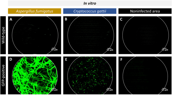
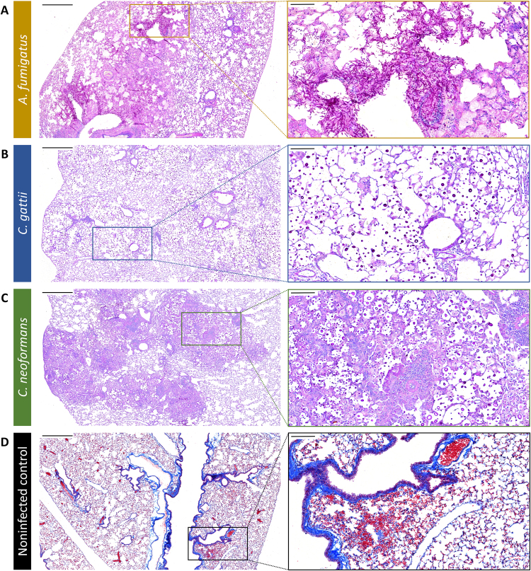
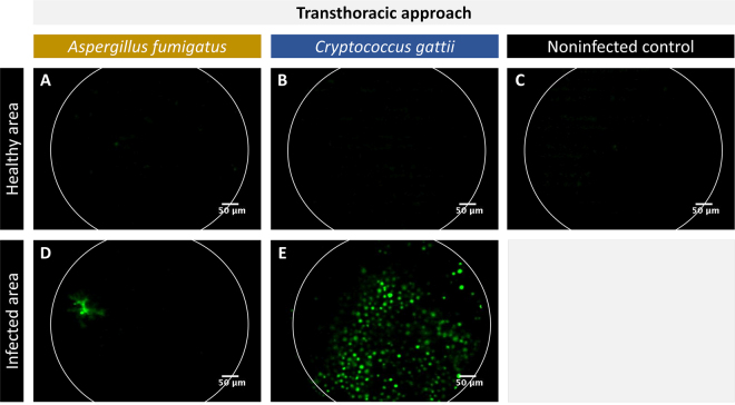
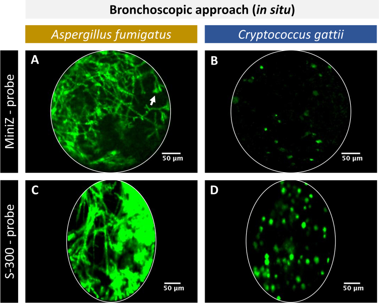
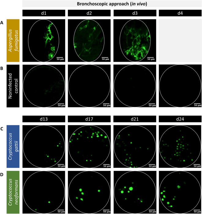
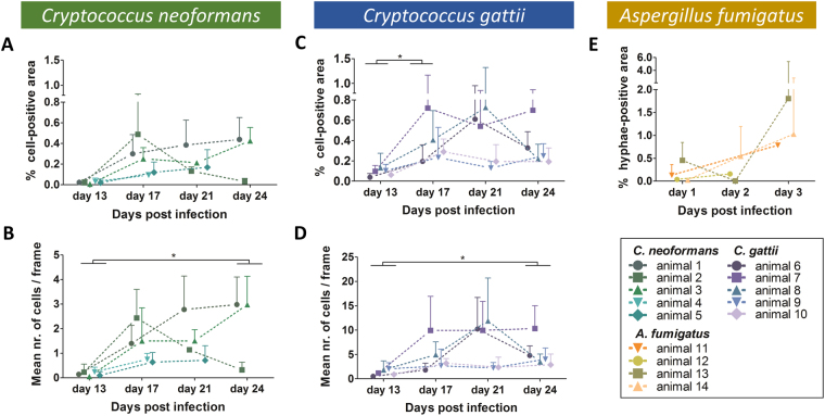
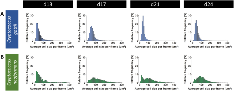
Respiratory diseases, such as pulmonary infections, are an important cause of morbidity and mortality worldwide. Preclinical studies often require invasive techniques to evaluate the extent of infection. Fibered confocal fluorescence microscopy (FCFM) is an emerging optical imaging technique that allows for real-time detection of fluorescently labeled cells within live animals, thereby bridging the gap between in vivo whole-body imaging methods and traditional histological examinations. Previously, the use of FCFM in preclinical lung research was limited to endpoint observations due to the invasive procedures required to access lungs. Here, we introduce a bronchoscopic FCFM approach that enabled in vivo visualization and morphological characterisation of fungal cells within lungs of mice suffering from pulmonary Aspergillus or Cryptococcus infections. The minimally invasive character of this approach allowed longitudinal monitoring of infection in free-breathing animals, thereby providing both visual and quantitative information on infection progression. Both the sensitivity and specificity of this technique were high during advanced stages of infection, allowing clear distinction between infected and non-infected animals. In conclusion, our study demonstrates the potential of this novel bronchoscopic FCFM approach to study pulmonary diseases, which can lead to novel insights in disease pathogenesis by allowing longitudinal in vivo microscopic examinations of the lungs.

摘要

呼吸系统疾病,如肺部感染,是全球发病率和死亡率的重要原因。临床前研究通常需要使用侵入性技术来评估感染的程度。纤维共聚焦荧光显微镜(FCFM)是一种新兴的光学成像技术,可实时检测活体动物内荧光标记的细胞,从而弥合体内全身成像方法与传统组织学检查之间的差距。以前,由于需要进行侵入性程序才能进入肺部,因此 FCFM 在临床前肺部研究中的应用仅限于终点观察。在这里,我们介绍了一种支气管 FCFM 方法,该方法能够在患有肺部曲霉菌或隐球菌感染的小鼠的肺部中可视化和形态学特征化真菌细胞。这种方法的微创性允许对自由呼吸的动物进行纵向监测感染,从而提供感染进展的视觉和定量信息。在感染的后期阶段,该技术具有较高的灵敏度和特异性,可清晰区分感染和未感染的动物。总之,我们的研究表明,这种新型支气管 FCFM 方法具有研究肺部疾病的潜力,通过允许对肺部进行纵向体内显微镜检查,可以为疾病发病机制提供新的见解。

https://cdn.ncbi.nlm.nih.gov/pmc/blobs/f3a8/5813038/8272396524ee/41598_2018_20545_Fig7_HTML.jpg
https://cdn.ncbi.nlm.nih.gov/pmc/blobs/f3a8/5813038/c16ddab5d722/41598_2018_20545_Fig1_HTML.jpg
https://cdn.ncbi.nlm.nih.gov/pmc/blobs/f3a8/5813038/75d4bc784c52/41598_2018_20545_Fig2_HTML.jpg
https://cdn.ncbi.nlm.nih.gov/pmc/blobs/f3a8/5813038/21b2b1acfa24/41598_2018_20545_Fig3_HTML.jpg
https://cdn.ncbi.nlm.nih.gov/pmc/blobs/f3a8/5813038/0ca235e08209/41598_2018_20545_Fig4_HTML.jpg
https://cdn.ncbi.nlm.nih.gov/pmc/blobs/f3a8/5813038/d7f029360dc0/41598_2018_20545_Fig5_HTML.jpg
https://cdn.ncbi.nlm.nih.gov/pmc/blobs/f3a8/5813038/077274395544/41598_2018_20545_Fig6_HTML.jpg
https://cdn.ncbi.nlm.nih.gov/pmc/blobs/f3a8/5813038/8272396524ee/41598_2018_20545_Fig7_HTML.jpg

相似文献

1
Bronchoscopic fibered confocal fluorescence microscopy for longitudinal in vivo assessment of pulmonary fungal infections in free-breathing mice.支气管镜纤维共聚焦荧光显微镜用于自由呼吸小鼠肺部真菌感染的纵向体内评估。
Sci Rep. 2018 Feb 14;8(1):3009. doi: 10.1038/s41598-018-20545-4.
2
In vivo molecular microimaging of pulmonary aspergillosis.肺曲霉病的体内分子显微成像
Med Mycol. 2013 May;51(4):352-60. doi: 10.3109/13693786.2012.729138. Epub 2012 Oct 23.
3
In vivo and in situ imaging of experimental invasive pulmonary aspergillosis using fibered confocal fluorescence microscopy.使用纤维共聚焦荧光显微镜对实验性侵袭性肺曲霉病进行体内和原位成像。
Med Mycol. 2012 May;50(4):386-95. doi: 10.3109/13693786.2011.617788. Epub 2011 Oct 17.
4
Longitudinal, in vivo assessment of invasive pulmonary aspergillosis in mice by computed tomography and magnetic resonance imaging.通过计算机断层扫描和磁共振成像对小鼠侵袭性肺曲霉病进行纵向活体评估。
Lab Invest. 2016 Jun;96(6):692-704. doi: 10.1038/labinvest.2016.45. Epub 2016 Mar 28.
5
Detection of Aspergillus fumigatus pulmonary fungal infections in mice with (99m)Tc-labeled MORF oligomers targeting ribosomal RNA.(99m)Tc 标记的针对核糖体 RNA 的 MORF 寡聚物检测小鼠中的烟曲霉肺部真菌感染。
Nucl Med Biol. 2013 Jan;40(1):89-96. doi: 10.1016/j.nucmedbio.2012.10.001. Epub 2012 Nov 9.
6
In vivo imaging of the airway wall in asthma: fibered confocal fluorescence microscopy in relation to histology and lung function.哮喘气道壁的体内成像:纤维共聚焦荧光显微镜与组织学和肺功能的关系。
Respir Res. 2011 Jun 23;12(1):85. doi: 10.1186/1465-9921-12-85.
7
A Multimodal Imaging Approach Enables Assessment of Antifungal Treatment in a Mouse Model of Invasive Pulmonary Aspergillosis.多模态影像学方法可用于评估侵袭性肺曲霉病小鼠模型中的抗真菌治疗效果。
Antimicrob Agents Chemother. 2018 Jun 26;62(7). doi: 10.1128/AAC.00240-18. Print 2018 Jul.
8
In vivo imaging of the bronchial wall microstructure using fibered confocal fluorescence microscopy.使用纤维共聚焦荧光显微镜对支气管壁微观结构进行体内成像。
Am J Respir Crit Care Med. 2007 Jan 1;175(1):22-31. doi: 10.1164/rccm.200605-684OC. Epub 2006 Oct 5.
9
Bioluminescence Imaging, a Powerful Tool to Assess Fungal Burden in Live Mouse Models of Infection.生物发光成像技术,一种用于评估活体感染小鼠模型中真菌负担的强大工具。
Methods Mol Biol. 2023;2667:197-210. doi: 10.1007/978-1-0716-3199-7_15.
10
Cyclophosphamide reduces dectin-1 expression in the lungs of naive and Aspergillus fumigatus-infected mice.环磷酰胺可降低未感染和烟曲霉感染小鼠肺部的 dectin-1 表达。
Med Mycol. 2010 Mar;48(2):303-9. doi: 10.1080/13693780903136887.

引用本文的文献

1
Fungal infections in focus: accelerating non-invasive imaging from preclinical insights to clinical breakthroughs.聚焦真菌感染:加速非侵入性成像从临床前洞察到临床突破的进程。
Npj Imaging. 2025 Sep 16;3(1):42. doi: 10.1038/s44303-025-00105-y.
2
Quantitative MRI of a Cerebral Cryptococcoma Mouse Model for In Vivo Distinction between Different Cryptococcal Molecular Types.用于体内区分不同隐球菌分子类型的脑隐球菌瘤小鼠模型的定量磁共振成像
J Fungi (Basel). 2024 Aug 22;10(8):593. doi: 10.3390/jof10080593.
3
Preclinical Models for Cryptococcosis of the CNS and Their Characterization Using In Vivo Imaging Techniques.

本文引用的文献

1
Searching for the Optimal Fluorophore to Label Antimicrobial Peptides.寻找用于标记抗菌肽的最佳荧光团。
ACS Comb Sci. 2016 Nov 14;18(11):689-696. doi: 10.1021/acscombsci.6b00081. Epub 2016 Oct 20.
2
Usefulness of Bronchoscopic Probe-Based Confocal Laser Endomicroscopy in the Diagnosis of Pneumocystis jirovecii Pneumonia.基于支气管镜探头的共聚焦激光显微内镜检查在诊断耶氏肺孢子菌肺炎中的应用价值
Respiration. 2016;92(1):40-7. doi: 10.1159/000447431. Epub 2016 Jul 1.
3
Longitudinal, in vivo assessment of invasive pulmonary aspergillosis in mice by computed tomography and magnetic resonance imaging.
中枢神经系统隐球菌病的临床前模型及其使用体内成像技术的表征
J Fungi (Basel). 2024 Feb 12;10(2):146. doi: 10.3390/jof10020146.
4
The role of confocal laser endomicroscopy in pulmonary medicine.共聚焦激光内镜在肺部医学中的应用。
Eur Respir Rev. 2023 Jan 25;32(167). doi: 10.1183/16000617.0185-2022. Print 2023 Mar 31.
5
Many ways, one microorganism: Several approaches to study Malassezia in interactions with model hosts.多种方法,一种微生物:几种研究马拉色菌与模型宿主相互作用的方法。
PLoS Pathog. 2022 Sep 8;18(9):e1010784. doi: 10.1371/journal.ppat.1010784. eCollection 2022 Sep.
6
Fluorescent toys 'n' tools lighting the way in fungal research.荧光玩具和工具照亮真菌研究之路。
FEMS Microbiol Rev. 2021 Sep 8;45(5). doi: 10.1093/femsre/fuab013.
7
From Mouse to Man and Back: Closing the Correlation Gap between Imaging and Histopathology for Lung Diseases.从小鼠到人类再回归:弥合肺部疾病影像学与组织病理学之间的关联差距
Diagnostics (Basel). 2020 Aug 26;10(9):636. doi: 10.3390/diagnostics10090636.
8
Redundant Trojan horse and endothelial-circulatory mechanisms for host-mediated spread of Candida albicans yeast.冗余的特洛伊木马和内皮循环机制促进白念珠菌酵母的宿主介导传播。
PLoS Pathog. 2020 Aug 10;16(8):e1008414. doi: 10.1371/journal.ppat.1008414. eCollection 2020 Aug.
9
Analysis of factors of pulmonary fungal infection in mice in respiratory medicine department based on logistic regression analysis model and Progranulin.基于逻辑回归分析模型和颗粒蛋白前体对呼吸内科小鼠肺部真菌感染因素的分析
Saudi J Biol Sci. 2020 Feb;27(2):629-635. doi: 10.1016/j.sjbs.2019.12.001. Epub 2019 Dec 17.
10
Illuminating Macrophage Contributions to Host-Pathogen Interactions : the Power of Zebrafish.阐明巨噬细胞在宿主-病原体相互作用中的贡献:斑马鱼的力量。
Infect Immun. 2020 Jun 22;88(7). doi: 10.1128/IAI.00906-19.
通过计算机断层扫描和磁共振成像对小鼠侵袭性肺曲霉病进行纵向活体评估。
Lab Invest. 2016 Jun;96(6):692-704. doi: 10.1038/labinvest.2016.45. Epub 2016 Mar 28.
4
Spacer-free BODIPY fluorogens in antimicrobial peptides for direct imaging of fungal infection in human tissue.用于人体组织真菌感染直接成像的抗菌肽中无间隔臂的BODIPY荧光团。
Nat Commun. 2016 Mar 9;7:10940. doi: 10.1038/ncomms10940.
5
Longitudinal micro-CT provides biomarkers of lung disease that can be used to assess the effect of therapy in preclinical mouse models, and reveal compensatory changes in lung volume.纵向微型计算机断层扫描提供了肺部疾病的生物标志物,可用于评估临床前小鼠模型中的治疗效果,并揭示肺容积的代偿性变化。
Dis Model Mech. 2016 Jan;9(1):91-8. doi: 10.1242/dmm.020321. Epub 2015 Nov 12.
6
A Novel Method for In Vivo Imaging of Solitary Lung Nodules Using Navigational Bronchoscopy and Confocal Laser Microendoscopy.一种使用导航支气管镜和共聚焦激光显微内镜对孤立性肺结节进行体内成像的新方法。
Lung. 2015 Oct;193(5):773-8. doi: 10.1007/s00408-015-9769-8. Epub 2015 Jul 28.
7
Comparison of in vivo probe-based confocal laser endomicroscopy with histopathology in lung cancer: A move toward optical biopsy.肺癌中基于探头的共聚焦激光内镜检查与组织病理学的体内比较:迈向光学活检的一步。
Respirology. 2015 Aug;20(6):967-74. doi: 10.1111/resp.12578. Epub 2015 Jun 19.
8
Real-time in-vivo imaging of pulmonary capillary perfusion using probe-based confocal laser scanning endomicroscopy in pigs: An interventional laboratory study.使用基于探头的共聚焦激光扫描内镜对猪肺毛细血管灌注进行实时体内成像:一项介入性实验室研究。
Eur J Anaesthesiol. 2015 Jun;32(6):392-9. doi: 10.1097/EJA.0000000000000260.
9
In vivo fluorescence microscopy: lessons from observing cell behavior in their native environment.体内荧光显微镜检查:观察细胞在其天然环境中的行为所获得的经验教训。
Physiology (Bethesda). 2015 Jan;30(1):40-9. doi: 10.1152/physiol.00019.2014.
10
The case of diagnostics of invasive pulmonary aspergillosis by in vivo probe-based confocal laser endomicroscopy of central and distal airways.通过基于体内探头的共聚焦激光内镜检查对中央和远端气道进行侵袭性肺曲霉病诊断的病例
Med Mycol Case Rep. 2014 Jul 24;5:35-9. doi: 10.1016/j.mmcr.2014.07.004. eCollection 2014 Jul.