• 文献检索
  • 文档翻译
  • 深度研究
  • 学术资讯
  • Suppr Zotero 插件Zotero 插件
  • 邀请有礼
  • 套餐&价格
  • 历史记录
应用&插件
Suppr Zotero 插件Zotero 插件浏览器插件Mac 客户端Windows 客户端微信小程序
定价
高级版会员购买积分包购买API积分包
服务
文献检索文档翻译深度研究API 文档MCP 服务
关于我们
关于 Suppr公司介绍联系我们用户协议隐私条款
关注我们

Suppr 超能文献

核心技术专利:CN118964589B侵权必究
粤ICP备2023148730 号-1Suppr @ 2026

文献检索

告别复杂PubMed语法,用中文像聊天一样搜索,搜遍4000万医学文献。AI智能推荐,让科研检索更轻松。

立即免费搜索

文件翻译

保留排版,准确专业,支持PDF/Word/PPT等文件格式,支持 12+语言互译。

免费翻译文档

深度研究

AI帮你快速写综述,25分钟生成高质量综述,智能提取关键信息,辅助科研写作。

立即免费体验

使用正常和青光眼眼睛的体内图像进行3D建模,以表征筛板表面和孔隙几何结构。

3D modeling to characterize lamina cribrosa surface and pore geometries using in vivo images from normal and glaucomatous eyes.

作者信息

Sredar Nripun, Ivers Kevin M, Queener Hope M, Zouridakis George, Porter Jason

机构信息

Department of Computer Science, University of Houston, Houston, TX 77004, USA.

出版信息

Biomed Opt Express. 2013 Jun 14;4(7):1153-65. doi: 10.1364/BOE.4.001153. Print 2013 Jul 1.

DOI:10.1364/BOE.4.001153
PMID:23847739
原文链接:https://pmc.ncbi.nlm.nih.gov/articles/PMC3704095/
Abstract

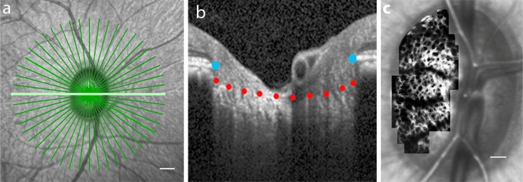
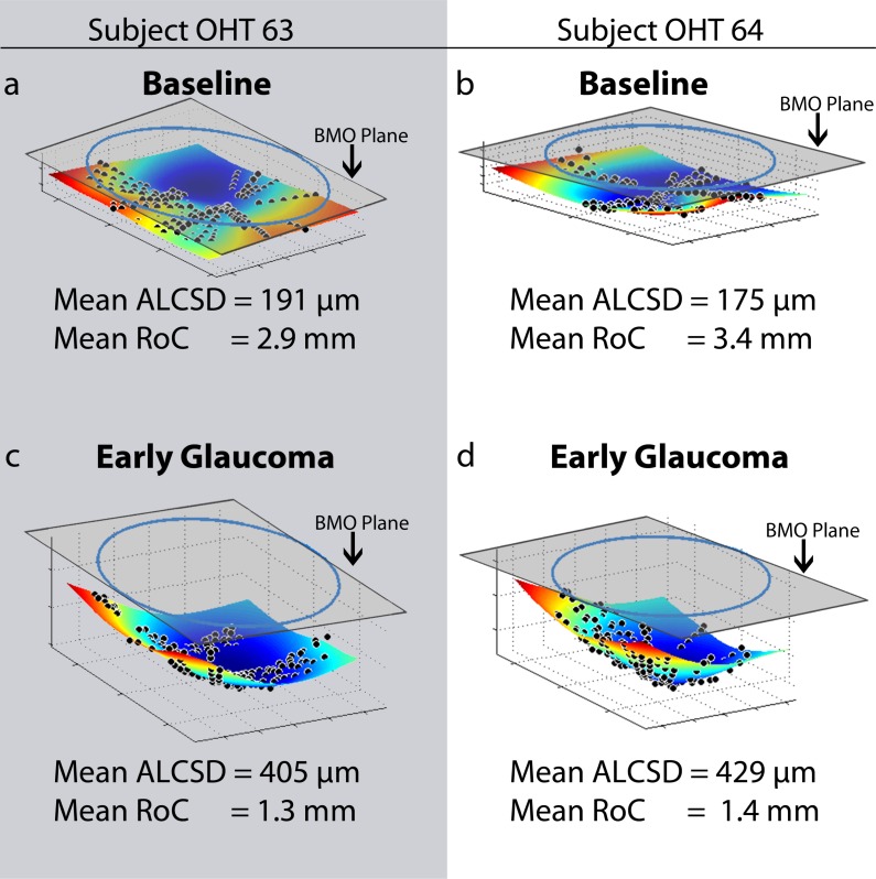
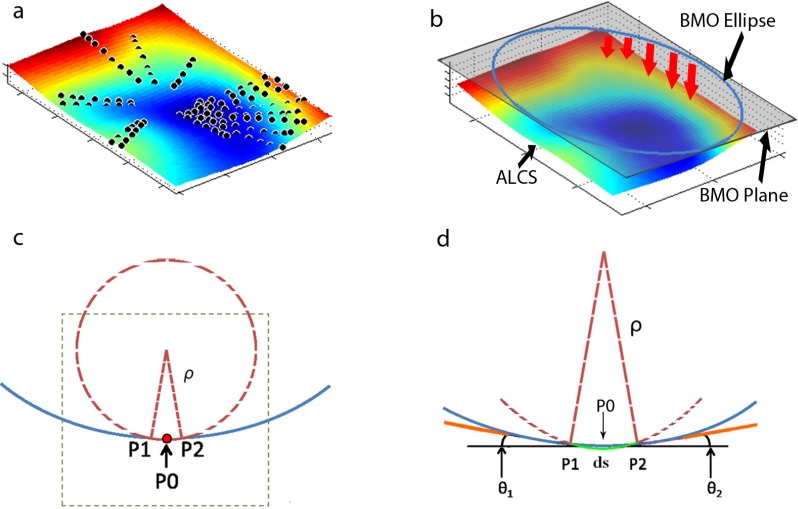
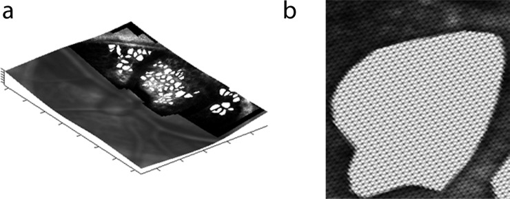
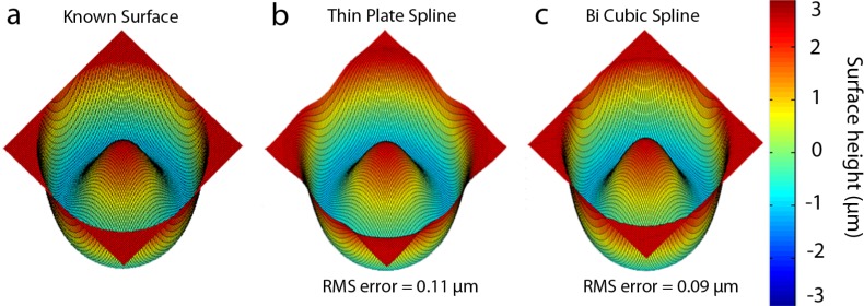
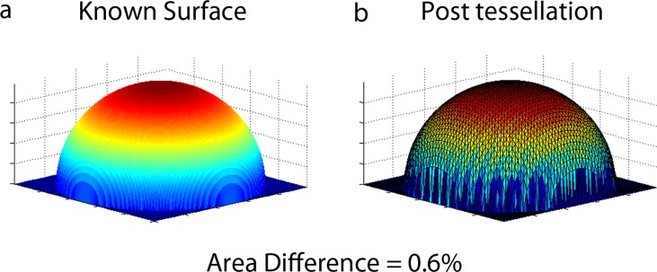
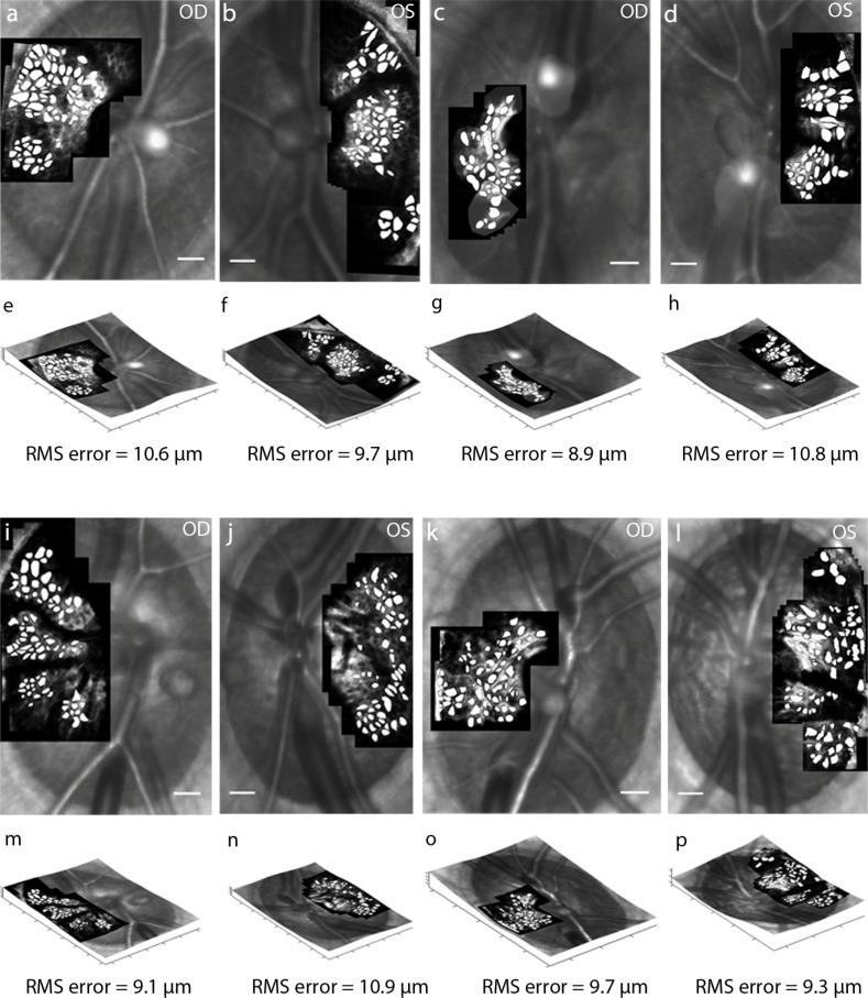
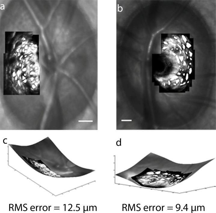
En face adaptive optics scanning laser ophthalmoscope (AOSLO) images of the anterior lamina cribrosa surface (ALCS) represent a 2D projected view of a 3D laminar surface. Using spectral domain optical coherence tomography images acquired in living monkey eyes, a thin plate spline was used to model the ALCS in 3D. The 2D AOSLO images were registered and projected onto the 3D surface that was then tessellated into a triangular mesh to characterize differences in pore geometry between 2D and 3D images. Following 3D transformation of the anterior laminar surface in 11 normal eyes, mean pore area increased by 5.1 ± 2.0% with a minimal change in pore elongation (mean change = 0.0 ± 0.2%). These small changes were due to the relatively flat laminar surfaces inherent in normal eyes (mean radius of curvature = 3.0 ± 0.5 mm). The mean increase in pore area was larger following 3D transformation in 4 glaucomatous eyes (16.2 ± 6.0%) due to their more steeply curved laminar surfaces (mean radius of curvature = 1.3 ± 0.1 mm), while the change in pore elongation was comparable to that in normal eyes (-0.2 ± 2.0%). This 3D transformation and tessellation method can be used to better characterize and track 3D changes in laminar pore and surface geometries in glaucoma.

摘要

筛板前表面(ALCS)的正面自适应光学扫描激光检眼镜(AOSLO)图像代表三维层状表面的二维投影视图。利用在活体猴眼中获取的光谱域光学相干断层扫描图像,使用薄板样条对ALCS进行三维建模。将二维AOSLO图像配准并投影到三维表面上,然后将该表面细分为三角形网格,以表征二维和三维图像之间孔隙几何形状的差异。在对11只正常眼睛的前层状表面进行三维变换后,平均孔隙面积增加了5.1±2.0%,孔隙伸长率变化最小(平均变化=0.0±0.2%)。这些微小变化是由于正常眼睛固有的相对平坦的层状表面(平均曲率半径=3.0±0.5毫米)。在4只青光眼眼睛中进行三维变换后,平均孔隙面积增加更大(16.2±6.0%),这是因为它们的层状表面弯曲更陡峭(平均曲率半径=1.3±0.1毫米),而孔隙伸长率的变化与正常眼睛相当(-0.2±2.0%)。这种三维变换和细分方法可用于更好地表征和跟踪青光眼中层状孔隙和表面几何形状的三维变化。

https://cdn.ncbi.nlm.nih.gov/pmc/blobs/56e4/3704095/0a72044b7757/boe-4-7-1153-g008.jpg
https://cdn.ncbi.nlm.nih.gov/pmc/blobs/56e4/3704095/c9557f483830/boe-4-7-1153-g001.jpg
https://cdn.ncbi.nlm.nih.gov/pmc/blobs/56e4/3704095/ce78c46344ff/boe-4-7-1153-g002.jpg
https://cdn.ncbi.nlm.nih.gov/pmc/blobs/56e4/3704095/b1de801c53c0/boe-4-7-1153-g003.jpg
https://cdn.ncbi.nlm.nih.gov/pmc/blobs/56e4/3704095/397c5736d57d/boe-4-7-1153-g004.jpg
https://cdn.ncbi.nlm.nih.gov/pmc/blobs/56e4/3704095/13d128fb57de/boe-4-7-1153-g005.jpg
https://cdn.ncbi.nlm.nih.gov/pmc/blobs/56e4/3704095/3ffc29f3bcc0/boe-4-7-1153-g006.jpg
https://cdn.ncbi.nlm.nih.gov/pmc/blobs/56e4/3704095/a2a4bdca6ae2/boe-4-7-1153-g007.jpg
https://cdn.ncbi.nlm.nih.gov/pmc/blobs/56e4/3704095/0a72044b7757/boe-4-7-1153-g008.jpg

相似文献

1
3D modeling to characterize lamina cribrosa surface and pore geometries using in vivo images from normal and glaucomatous eyes.使用正常和青光眼眼睛的体内图像进行3D建模,以表征筛板表面和孔隙几何结构。
Biomed Opt Express. 2013 Jun 14;4(7):1153-65. doi: 10.1364/BOE.4.001153. Print 2013 Jul 1.
2
In Vivo Changes in Lamina Cribrosa Microarchitecture and Optic Nerve Head Structure in Early Experimental Glaucoma.早期实验性青光眼中筛板微结构和视神经乳头结构的体内变化
PLoS One. 2015 Jul 31;10(7):e0134223. doi: 10.1371/journal.pone.0134223. eCollection 2015.
3
Reproducibility of measuring lamina cribrosa pore geometry in human and nonhuman primates with in vivo adaptive optics imaging.利用活体自适应光学成像技术测量人类和非人类灵长类动物视乳头筛板孔几何形状的可重复性。
Invest Ophthalmol Vis Sci. 2011 Jul 23;52(8):5473-80. doi: 10.1167/iovs.11-7347.
4
In vivo imaging of lamina cribrosa pores by adaptive optics scanning laser ophthalmoscopy.自适应光学扫描激光检眼镜对神经纤维层筛板孔的活体成像。
Invest Ophthalmol Vis Sci. 2012 Jun 26;53(7):4111-9. doi: 10.1167/iovs.11-7536.
5
Adaptive optics scanning laser ophthalmoscopy for in vivo imaging of lamina cribrosa.用于筛板体内成像的自适应光学扫描激光检眼镜
J Opt Soc Am A Opt Image Sci Vis. 2007 May;24(5):1417-25. doi: 10.1364/josaa.24.001417.
6
Deformation of the lamina cribrosa and anterior scleral canal wall in early experimental glaucoma.早期实验性青光眼中筛板和前巩膜管管壁的变形
Invest Ophthalmol Vis Sci. 2003 Feb;44(2):623-37. doi: 10.1167/iovs.01-1282.
7
Repeatability of in vivo 3D lamina cribrosa microarchitecture using adaptive optics spectral domain optical coherence tomography.使用自适应光学光谱域光学相干断层扫描技术对体内三维筛板微结构的可重复性研究
Biomed Opt Express. 2014 Mar 10;5(4):1114-23. doi: 10.1364/BOE.5.001114. eCollection 2014 Apr 1.
8
Three-dimensional high-speed optical coherence tomography imaging of lamina cribrosa in glaucoma.青光眼筛板的三维高速光学相干断层扫描成像
Ophthalmology. 2009 Feb;116(2):214-22. doi: 10.1016/j.ophtha.2008.09.008. Epub 2008 Dec 16.
9
Optic Nerve Head Deformation in Glaucoma: A Prospective Analysis of Optic Nerve Head Surface and Lamina Cribrosa Surface Displacement.青光眼视乳头变形:视乳头表面和神经纤维层表面位移的前瞻性分析。
Ophthalmology. 2015 Jul;122(7):1317-29. doi: 10.1016/j.ophtha.2015.02.035. Epub 2015 May 9.
10
In vivo morphometry of the lamina cribrosa and its relation to visual field loss in glaucoma.青光眼患者筛板的活体形态测量及其与视野缺损的关系。
Curr Eye Res. 1998 Apr;17(4):363-9. doi: 10.1080/02713689808951216.

引用本文的文献

1
Lamina Cribrosa Insertions Into the Sclera Are Sparser, Narrower, and More Slanted in the Anterior Lamina.前层视乳头筛板插入巩膜处的板层插入物更稀疏、更狭窄且更倾斜。
Invest Ophthalmol Vis Sci. 2024 Apr 1;65(4):35. doi: 10.1167/iovs.65.4.35.
2
The Curvature, Collagen Network Structure, and Their Relationship to the Pressure-Induced Strain Response of the Human Lamina Cribrosa in Normal and Glaucoma Eyes.正常眼和青光眼眼中人板层 cribrosa 的曲率、胶原网络结构及其与眼压诱导应变响应的关系。
J Biomech Eng. 2023 Oct 1;145(10). doi: 10.1115/1.4062846.
3
Longitudinal In Vivo Changes in Radial Peripapillary Capillaries and Optic Nerve Head Structure in Non-Human Primates With Early Experimental Glaucoma.

本文引用的文献

1
In vivo imaging of lamina cribrosa pores by adaptive optics scanning laser ophthalmoscopy.自适应光学扫描激光检眼镜对神经纤维层筛板孔的活体成像。
Invest Ophthalmol Vis Sci. 2012 Jun 26;53(7):4111-9. doi: 10.1167/iovs.11-7536.
2
Three-dimensional evaluation of the lamina cribrosa using spectral-domain optical coherence tomography in glaucoma.应用频域光学相干断层扫描对青光眼视盘筛板进行三维评估。
Invest Ophthalmol Vis Sci. 2012 Jan 20;53(1):198-204. doi: 10.1167/iovs.11-7848.
3
Reproducibility of measuring lamina cribrosa pore geometry in human and nonhuman primates with in vivo adaptive optics imaging.
早期实验性青光眼非人类灵长类动物视乳头周围毛细血管和视神经头部结构的纵向活体变化。
Invest Ophthalmol Vis Sci. 2022 Jan 3;63(1):10. doi: 10.1167/iovs.63.1.10.
4
The optic nerve head, lamina cribrosa, and nerve fiber layer in non-myopic and myopic children.非近视和近视儿童的视神经头、筛板和神经纤维层。
Exp Eye Res. 2020 Jun;195:108041. doi: 10.1016/j.exer.2020.108041. Epub 2020 Apr 28.
5
Thin Lamina Cribrosa Beams Have Different Collagen Microstructure Than Thick Beams.薄视盘小梁梁比厚梁具有不同的胶原微结构。
Invest Ophthalmol Vis Sci. 2018 Sep 4;59(11):4653-4661. doi: 10.1167/iovs.18-24763.
6
Seeing the Hidden Lamina: Effects of Exsanguination on the Optic Nerve Head.观察潜在的神经层:失血对视神经头的影响。
Invest Ophthalmol Vis Sci. 2018 May 1;59(6):2564-2575. doi: 10.1167/iovs.17-23356.
7
Lamina Cribrosa Pore Shape and Size as Predictors of Neural Tissue Mechanical Insult.筛板孔的形状和大小作为神经组织机械损伤的预测指标
Invest Ophthalmol Vis Sci. 2017 Oct 1;58(12):5336-5346. doi: 10.1167/iovs.17-22015.
8
Review of adaptive optics OCT (AO-OCT): principles and applications for retinal imaging [Invited].自适应光学光学相干断层扫描(AO-OCT)综述:视网膜成像的原理与应用[特邀报告]
Biomed Opt Express. 2017 Apr 19;8(5):2536-2562. doi: 10.1364/BOE.8.002536. eCollection 2017 May 1.
9
Structural and Functional Evaluations for the Early Detection of Glaucoma.青光眼早期检测的结构和功能评估
Expert Rev Ophthalmol. 2016;11(5):367-376. doi: 10.1080/17469899.2016.1229599. Epub 2016 Sep 14.
10
A machine-learning graph-based approach for 3D segmentation of Bruch's membrane opening from glaucomatous SD-OCT volumes.基于机器学习图的方法用于从青光眼 SD-OCT 容积中分割布鲁赫膜开口的 3D 图像。
Med Image Anal. 2017 Jul;39:206-217. doi: 10.1016/j.media.2017.04.007. Epub 2017 May 6.
利用活体自适应光学成像技术测量人类和非人类灵长类动物视乳头筛板孔几何形状的可重复性。
Invest Ophthalmol Vis Sci. 2011 Jul 23;52(8):5473-80. doi: 10.1167/iovs.11-7347.
4
Longitudinal change detected by spectral domain optical coherence tomography in the optic nerve head and peripapillary retina in experimental glaucoma.实验性青光眼视盘和视盘周围视网膜的频域光学相干断层扫描检测到的纵向变化。
Invest Ophthalmol Vis Sci. 2011 Mar 2;52(3):1206-19. doi: 10.1167/iovs.10-5599.
5
A correction algorithm to simultaneously control dual deformable mirrors in a woofer-tweeter adaptive optics system.一种用于在低音-高音自适应光学系统中同时控制双变形镜的校正算法。
Opt Express. 2010 Aug 2;18(16):16671-84. doi: 10.1364/OE.18.016671.
6
Adaptive optics optical coherence tomography at 120,000 depth scans/s for non-invasive cellular phenotyping of the living human retina.用于活体人类视网膜非侵入性细胞表型分析的每秒120,000次深度扫描的自适应光学光学相干断层扫描技术
Opt Express. 2009 Oct 26;17(22):19382-400. doi: 10.1364/OE.17.019382.
7
Mechanical environment of the optic nerve head in glaucoma.青光眼视乳头的机械环境
Optom Vis Sci. 2008 Jun;85(6):425-35. doi: 10.1097/OPX.0b013e31817841cb.
8
3-D histomorphometry of the normal and early glaucomatous monkey optic nerve head: lamina cribrosa and peripapillary scleral position and thickness.正常及早期青光眼猴视神经乳头的三维组织形态计量学:筛板及视乳头周围巩膜的位置和厚度
Invest Ophthalmol Vis Sci. 2007 Oct;48(10):4597-607. doi: 10.1167/iovs.07-0349.
9
Adaptive optics scanning laser ophthalmoscopy for in vivo imaging of lamina cribrosa.用于筛板体内成像的自适应光学扫描激光检眼镜
J Opt Soc Am A Opt Image Sci Vis. 2007 May;24(5):1417-25. doi: 10.1364/josaa.24.001417.
10
Maximum permissible exposures for ocular safety (ANSI 2000), with emphasis on ophthalmic devices.眼部安全的最大允许暴露量(美国国家标准学会,2000年),重点关注眼科设备。
J Opt Soc Am A Opt Image Sci Vis. 2007 May;24(5):1250-65. doi: 10.1364/josaa.24.001250.