Department of Biomedical Engineering, Duke University, Durham, North Carolina, USA.
PLoS One. 2013 Jul 23;8(7):e68868. doi: 10.1371/journal.pone.0068868. Print 2013.
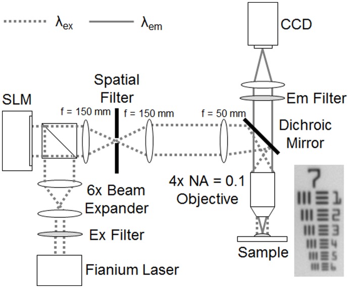
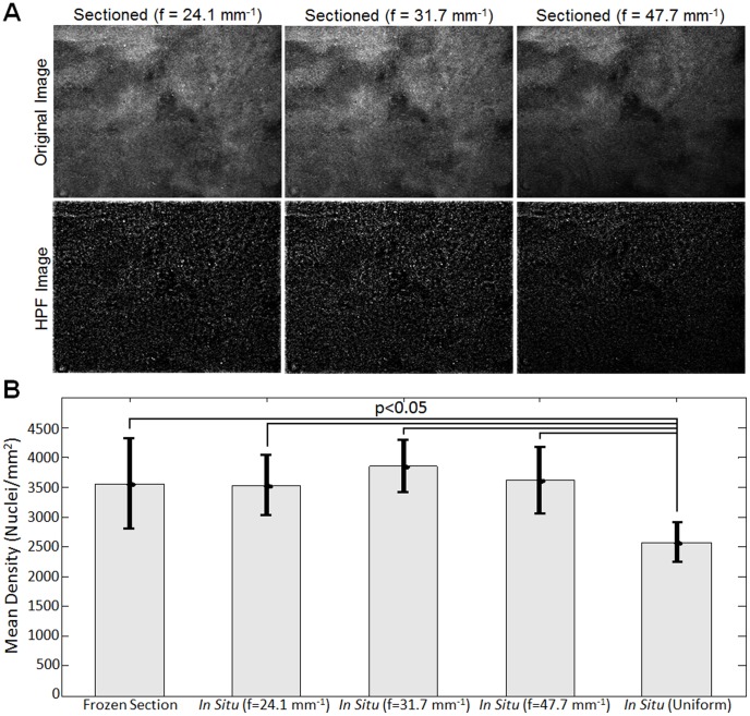
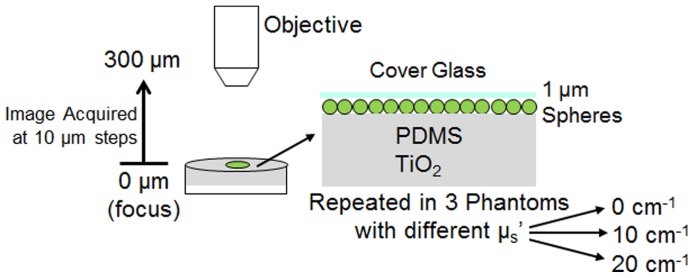
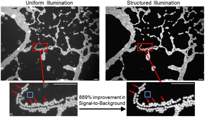
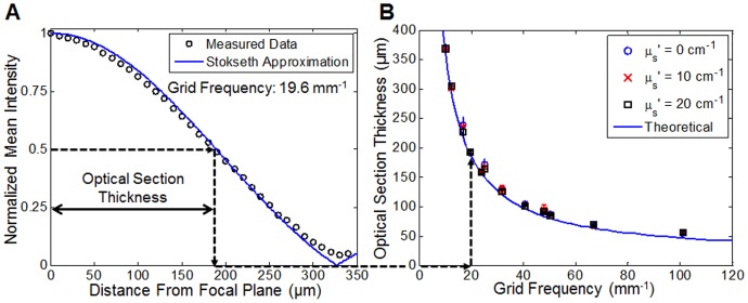
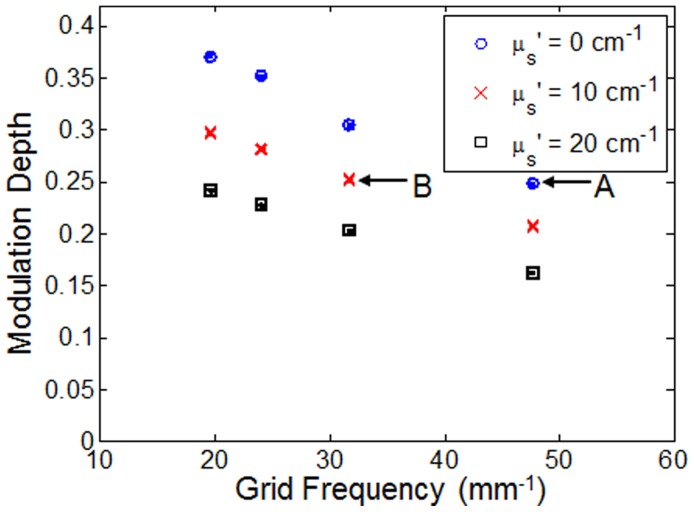
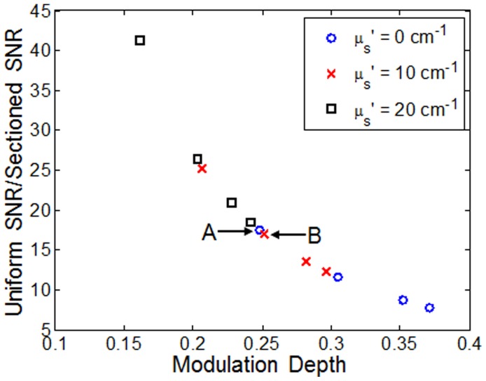
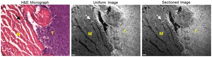
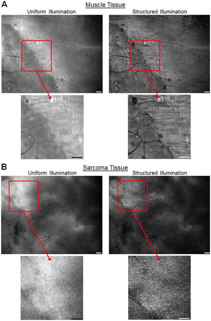
Cancer is associated with specific cellular morphological changes, such as increased nuclear size and crowding from rapidly proliferating cells. In situ tissue imaging using fluorescent stains may be useful for intraoperative detection of residual cancer in surgical tumor margins. We developed a widefield fluorescence structured illumination microscope (SIM) system with a single-shot FOV of 2.1 × 1.6 mm (3.4 mm(2)) and sub-cellular resolution (4.4 µm). The objectives of this work were to measure the relationship between illumination pattern frequency and optical sectioning strength and signal-to-noise ratio in turbid (i.e. thick) samples for selection of the optimum frequency, and to determine feasibility for detecting residual cancer on tumor resection margins, using a genetically engineered primary mouse model of sarcoma. The SIM system was tested in tissue mimicking solid phantoms with various scattering levels to determine impact of both turbidity and illumination frequency on two SIM metrics, optical section thickness and modulation depth. To demonstrate preclinical feasibility, ex vivo 50 µm frozen sections and fresh intact thick tissue samples excised from a primary mouse model of sarcoma were stained with acridine orange, which stains cell nuclei, skeletal muscle, and collagenous stroma. The cell nuclei were segmented using a high-pass filter algorithm, which allowed quantification of nuclear density. The results showed that the optimal illumination frequency was 31.7 µm(-1) used in conjunction with a 4 × 0.1 NA objective (v=0.165). This yielded an optical section thickness of 128 µm and an 8.9 × contrast enhancement over uniform illumination. We successfully demonstrated the ability to resolve cell nuclei in situ achieved via SIM, which allowed segmentation of nuclei from heterogeneous tissues in the presence of considerable background fluorescence. Specifically, we demonstrate that optical sectioning of fresh intact thick tissues performed equivalently in regards to nuclear density quantification, to physical frozen sectioning and standard microscopy.
癌症与特定的细胞形态变化有关,例如细胞核增大和快速增殖细胞的拥挤。使用荧光染色的原位组织成像可能有助于术中检测手术肿瘤边缘的残留癌症。我们开发了一种宽场荧光结构照明显微镜(SIM)系统,单次视野为 2.1×1.6mm(3.4mm²),具有亚细胞分辨率(4.4µm)。这项工作的目的是测量照明模式频率与混浊(即厚)样品中光学切片强度和信噪比之间的关系,以选择最佳频率,并使用肉瘤的遗传工程原发性小鼠模型确定检测肿瘤切除边缘残留癌症的可行性。SIM 系统在具有不同散射水平的组织模拟固体幻像中进行了测试,以确定浊度和照明频率对两个 SIM 指标,即光学切片厚度和调制深度的影响。为了证明临床前的可行性,使用吖啶橙对源自肉瘤的原发性小鼠模型的 50µm 冷冻切片和新鲜完整的厚组织样本进行了离体染色,吖啶橙可对细胞核、骨骼肌和胶原基质进行染色。使用高通滤波器算法对细胞核进行分割,允许对核密度进行定量。结果表明,最佳照明频率为 31.7µm(-1),与 4×0.1NA 物镜(v=0.165)结合使用。这产生了 128µm 的光学切片厚度和 8.9×的对比度增强,超过了均匀照明。我们成功地证明了通过 SIM 原位分辨细胞核的能力,这使得在存在相当大的背景荧光的情况下,能够对异质组织中的细胞核进行分割。具体而言,我们证明了新鲜完整的厚组织的光学切片在核密度定量方面与物理冷冻切片和标准显微镜相当。