Department of Anesthesiology, Medical College of Wisconsin, Milwaukee, Wisconsin, USA.
PLoS One. 2013 Jul 22;8(7):e70088. doi: 10.1371/journal.pone.0070088. Print 2013.
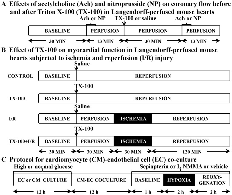
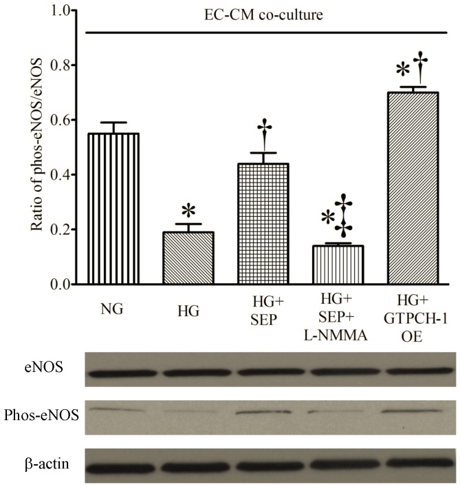
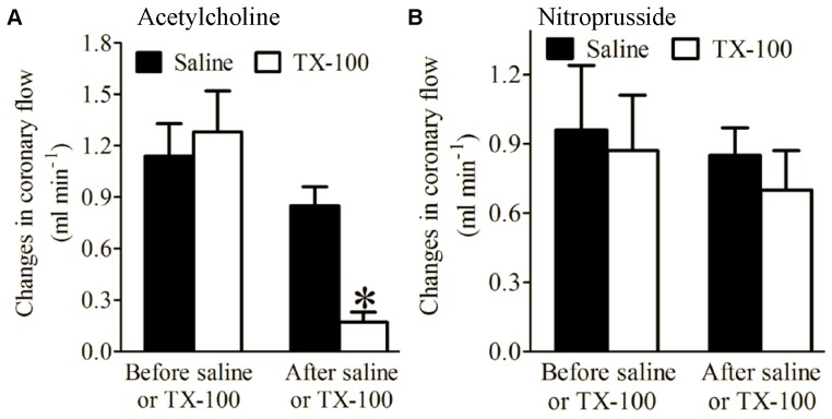
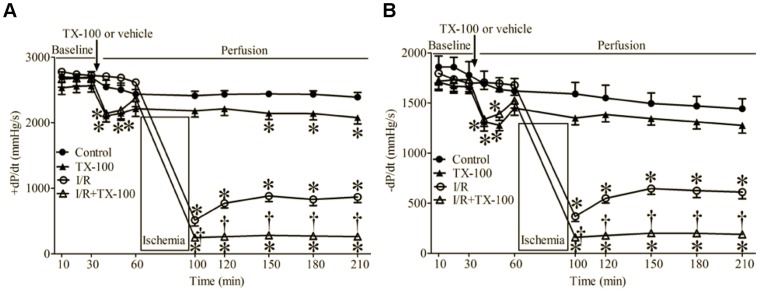
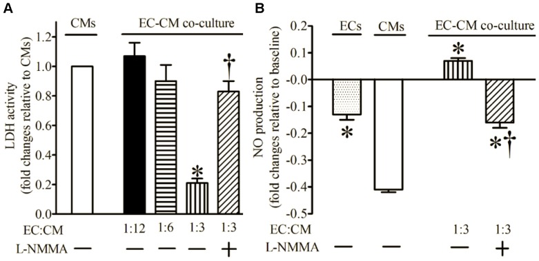
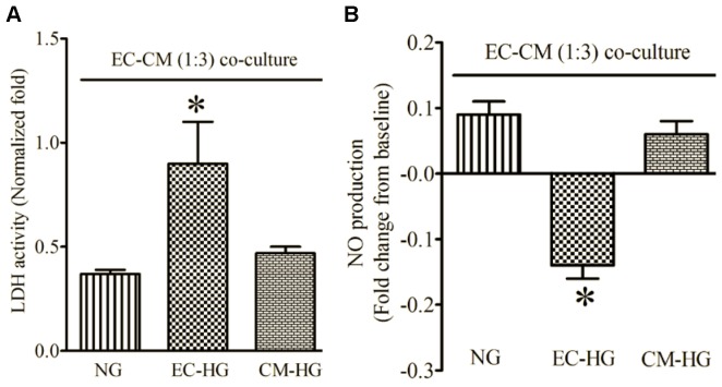
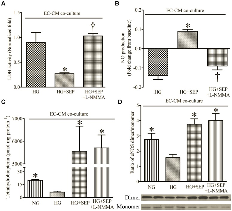
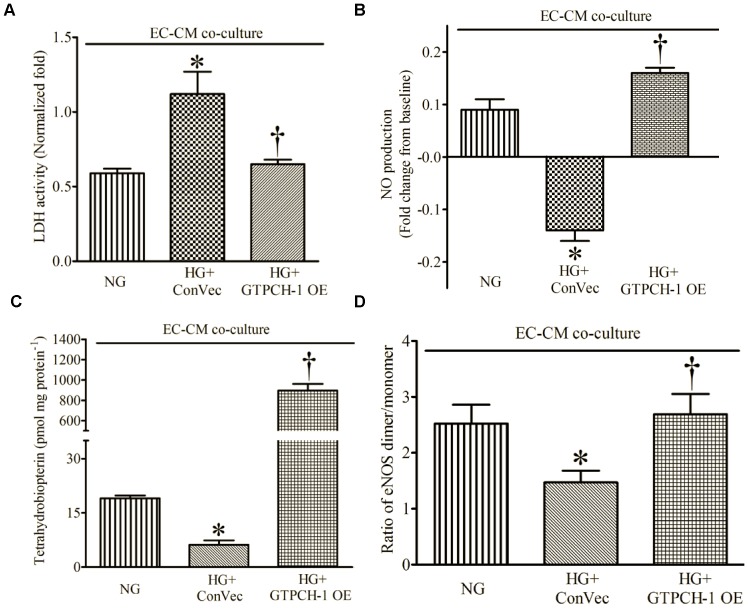
Endothelial-myocardial interactions may be critically important for ischemia/reperfusion injury. Tetrahydrobiopterin (BH4) is a required cofactor for nitric oxide (NO) production by endothelial NO synthase (eNOS). Hyperglycemia (HG) leads to significant increases in oxidative stress, oxidizing BH4 to enzymatically incompetent dihydrobiopterin. How alterations in endothelial BH4 content impact myocardial ischemia/reperfusion injury remains elusive. The aim of this study was to examine the effect of endothelial-myocardial interaction on ischemia/reperfusion injury, with an emphasis on the role of endothelial BH4 content. Langendorff-perfused mouse hearts were treated by triton X-100 to produce endothelial dysfunction and subsequently subjected to 30 min of ischemia followed by 2 h of reperfusion. The recovery of left ventricular systolic and diastolic function during reperfusion was impaired in triton X-100 treated hearts compared with vehicle-treated hearts. Cardiomyocytes (CMs) were co-cultured with endothelial cells (ECs) and subsequently subjected to 2 h of hypoxia followed by 2 h of reoxygenation. Addition of ECs to CMs at a ratio of 1∶3 significantly increased NO production and decreased lactate dehydrogenase activity compared with CMs alone. This EC-derived protection was abolished by HG. The addition of 100 µM sepiapterin (a BH4 precursor) or overexpression of GTP cyclohydrolase 1 (the rate-limiting enzyme for BH4 biosynthesis) in ECs by gene trasfer enhanced endothelial BH4 levels, the ratio of eNOS dimer/monomer, eNOS phosphorylation, and NO production and decreased lactate dehydrogenase activity in the presence of HG. These results demonstrate that increased BH4 content in ECs by either pharmacological or genetic approaches reduces myocardial damage during hypoxia/reoxygenation in the presence of HG. Maintaining sufficient endothelial BH4 is crucial for cardioprotection against hypoxia/reoxygenation injury.
内皮-心肌相互作用对于缺血/再灌注损伤可能至关重要。四氢生物蝶呤 (BH4) 是内皮型一氧化氮合酶 (eNOS) 产生一氧化氮 (NO) 的必需辅助因子。高血糖 (HG) 导致氧化应激显著增加,将 BH4 氧化为酶失活的二氢生物蝶呤。内皮 BH4 含量的变化如何影响心肌缺血/再灌注损伤仍不清楚。本研究旨在研究内皮-心肌相互作用对缺血/再灌注损伤的影响,重点关注内皮 BH4 含量的作用。用 Triton X-100 处理 Langendorff 灌注的小鼠心脏以产生内皮功能障碍,随后进行 30 分钟缺血,再灌注 2 小时。与载体处理的心脏相比,Triton X-100 处理的心脏在再灌注期间左心室收缩和舒张功能的恢复受损。与单独的心肌细胞 (CMs) 相比,将内皮细胞 (ECs) 与 CMs 共培养并随后进行 2 小时缺氧和 2 小时复氧,EC 向 CMs 的添加比例为 1∶3 显著增加了 NO 产生并降低了乳酸脱氢酶活性。这种 EC 衍生的保护作用被 HG 所消除。在 HG 存在的情况下,在 EC 中添加 100µM 蝶呤 (BH4 的前体) 或通过基因转移过表达 GTP 环化水解酶 1( BH4 生物合成的限速酶),可增强内皮细胞 BH4 水平、eNOS 二聚体/单体比、eNOS 磷酸化、NO 产生并降低乳酸脱氢酶活性。这些结果表明,通过药理学或遗传学方法增加 EC 中的 BH4 含量可减少 HG 存在时缺氧/复氧期间的心肌损伤。维持足够的内皮 BH4 对于对抗缺氧/复氧损伤的心脏保护至关重要。