Brown Ryan, Storey Pippa, Geppert Christian, McGorty KellyAnne, Leite Ana Paula Klautau, Babb James, Sodickson Daniel K, Wiggins Graham C, Moy Linda
Bernard and Irene Schwartz Center for Biomedical Imaging, Department of Radiology, New York University Langone Medical Center, 660 First Avenue, Room 401, New York, NY, USA,
Eur Radiol. 2013 Nov;23(11):2969-78. doi: 10.1007/s00330-013-2972-1. Epub 2013 Jul 30.
To evaluate the image quality of T1-weighted fat-suppressed breast MRI at 7 T and to compare 7-T and 3-T images.
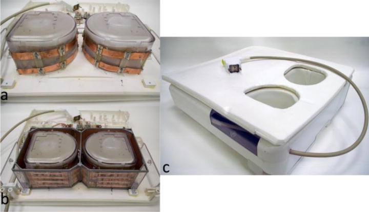
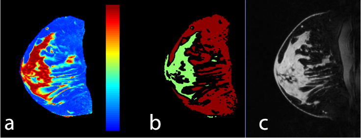
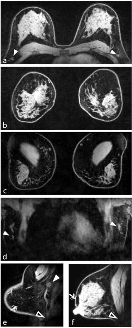
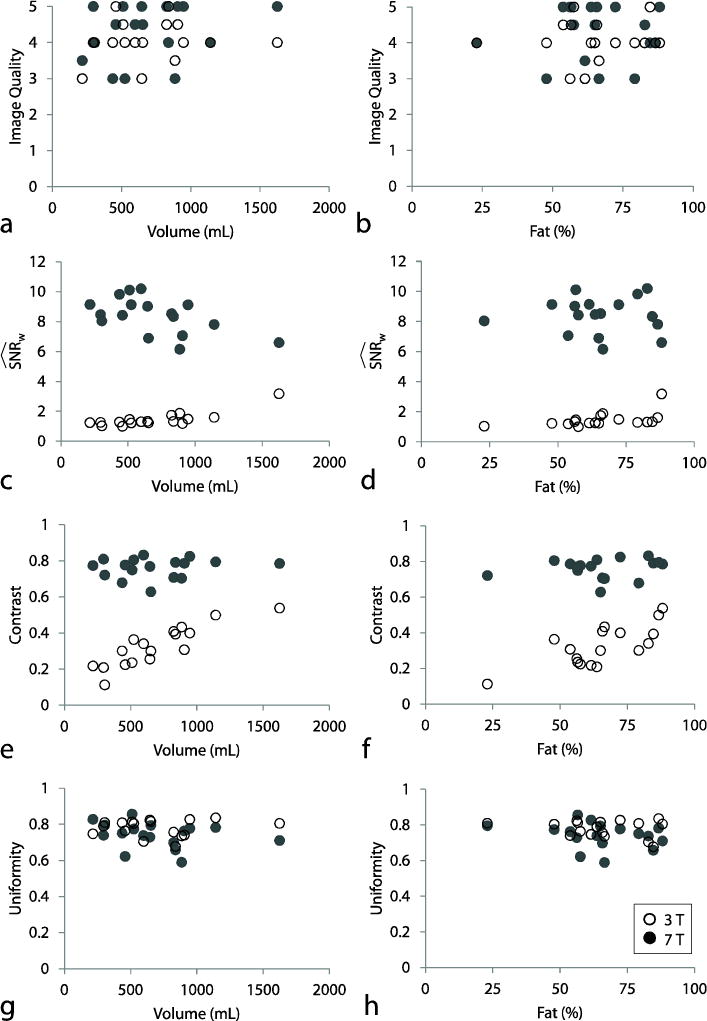
Seventeen subjects were imaged using a 7-T bilateral transmit-receive coil and 3D gradient echo sequence with adiabatic inversion-based fat suppression (FS). Images were graded on a five-point scale and quantitatively assessed through signal-to-noise ratio (SNR), fibroglandular/fat contrast and signal uniformity measurements.
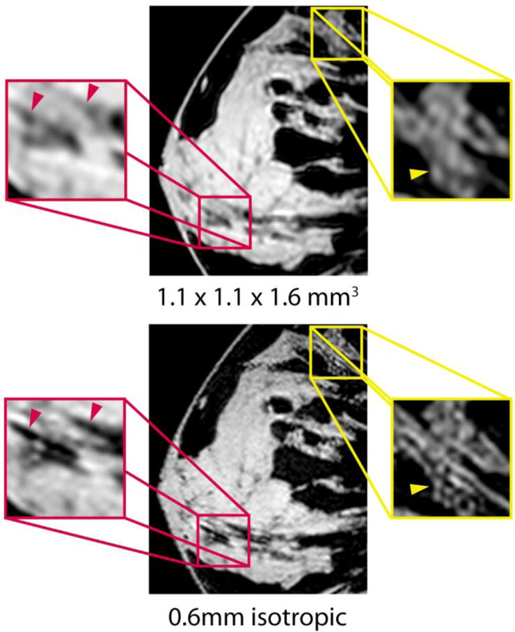
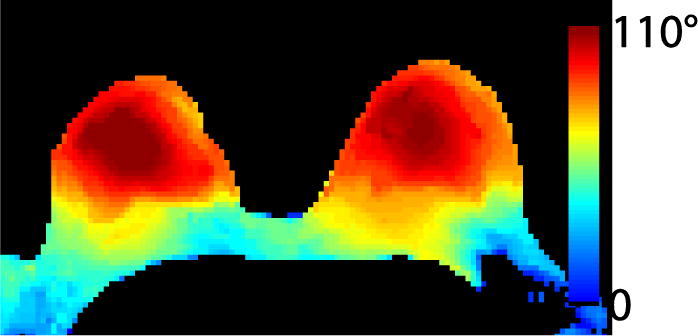
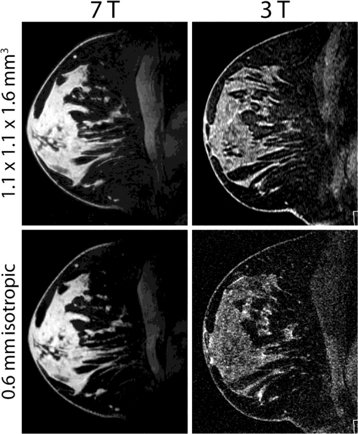
Image scores at 7 and 3 T were similar on standard-resolution images (1.1 × 1.1 × 1.1-1.6 mm(3)), indicating that high-quality breast imaging with clinical parameters can be performed at 7 T. The 7-T SNR advantage was underscored on 0.6-mm isotropic images, where image quality was significantly greater than at 3 T (4.2 versus 3.1, P ≤ 0.0001). Fibroglandular/fat contrast was more than two times higher at 7 T than at 3 T, owing to effective adiabatic inversion-based FS and the inherent 7-T signal advantage. Signal uniformity was comparable at 7 and 3 T (P < 0.05). Similar 7-T image quality was observed in all subjects, indicating robustness against anatomical variation.
The 7-T bilateral transmit-receive coil and adiabatic inversion-based FS technique produce image quality that is as good as or better than at 3 T.
• High image quality bilateral breast MRI is achievable with clinical parameters at 7 T. • 7-T high-resolution imaging improves delineation of subtle soft tissue structures. • Adiabatic-based fat suppression provides excellent fibroglandular/fat contrast at 7 T. • 7- and 3-T 3D T1-weighted gradient-echo images have similar signal uniformity. • The 7-T dual solenoid coil enables bilateral imaging without compromising uniformity.
评估7T场强下T1加权脂肪抑制乳腺MRI的图像质量,并比较7T和3T图像。
17名受试者使用7T双侧发射接收线圈和基于绝热反转脂肪抑制(FS)的3D梯度回波序列进行成像。图像按五分制评分,并通过信噪比(SNR)、纤维腺体/脂肪对比度和信号均匀性测量进行定量评估。
在标准分辨率图像(1.1×1.1×1.1-1.6mm³)上,7T和3T的图像评分相似,表明在7T场强下可利用临床参数进行高质量乳腺成像。在0.6mm各向同性图像上,7T的SNR优势更为突出,其图像质量显著高于3T(4.2对3.1, P≤0.0001)。由于有效的基于绝热反转的FS和7T固有的信号优势,7T时纤维腺体/脂肪对比度比3T时高出两倍多。7T和3T时的信号均匀性相当(P<0.05)。在所有受试者中均观察到相似的7T图像质量,表明其对解剖变异具有稳健性。
7T双侧发射接收线圈和基于绝热反转的FS技术产生的图像质量与3T时相当或更好。
• 在7T场强下利用临床参数可实现高质量双侧乳腺MRI成像。• 7T高分辨率成像可改善对细微软组织结构的描绘。• 基于绝热的脂肪抑制在7T时提供出色的纤维腺体/脂肪对比度。• 7T和3T的3D T1加权梯度回波图像具有相似的信号均匀性。• 7T双螺线管线圈可实现双侧成像而不影响均匀性。