Department of Radiology, Seoul National University College of Medicine, Seoul National University Hospital, and Institute of Radiation Medicine, Seoul 110-744, Korea.
Korean J Radiol. 2013 Jul-Aug;14(4):559-67. doi: 10.3348/kjr.2013.14.4.559. Epub 2013 Jul 17.
To evaluate the additional effect of sonoelastography on the radiologist's ability for distinguishing benign from malignant complex breast masses and to decide whether to perform biopsy by B-mode US.
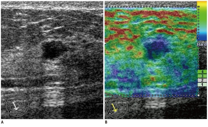
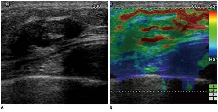
One hundred eighteen complex breast masses (15 malignant lesions, 103 benign lesions) were included. Five blinded readers independently assessed the likelihood of the malignancy score from 1 to 5 for two data sets (B-mode ultrasound alone and B-mode ultrasound with sonoelastography). Elasticity scores were categorized as 0, 1, or 2 based on the degree and distribution of strain of the echogenic component within complex masses. The readers were asked to downgrade the likelihood of the malignancy score when an elasticity score of 0 was assigned and to upgrade the likelihood of the malignancy score when an elasticity score of 2 was assigned. The likelihood of the malignancy score was maintained as it was for the lesions with an elasticity score of 1. The Az values, sensitivities, and specificities were compared.
The Az value of B-mode ultrasound with sonoelastography (mean, 0.863) was greater than that of B-mode ultrasound alone (mean, 0.731; p = 0.001-0.007) for all authors. The specificity of B-mode ultrasound with sonoelastography (mean, 37.1%) was greater than that of B-mode ultrasound alone (mean, 3.8%; p < 0.001) for all readers. The addition of sonoelastography led to changes in decisions. A mean of 33.6% of benign masses were recommended for follow-up instead of biopsy.
For complex breast masses, sonoelastography allows increase in both the accuracy in distinguishing benign from malignant lesions and the specificity in deciding whether to perform biopsy.
评估超声弹性成像对放射科医师鉴别良恶性复杂乳腺肿块的能力的额外作用,并决定是否进行 B 型超声引导下的活检。
纳入 118 个复杂乳腺肿块(15 个恶性病变,103 个良性病变)。5 位盲法读者分别对两个数据集(单纯 B 型超声和 B 型超声联合超声弹性成像)的恶性可能性评分从 1 到 5 进行评估。弹性评分根据复杂肿块内回声成分的应变程度和分布分为 0、1 或 2。当弹性评分为 0 时,读者被要求降低恶性评分的可能性,当弹性评分为 2 时,读者被要求提高恶性评分的可能性。当弹性评分为 1 时,病变的恶性评分保持不变。比较 Az 值、敏感度和特异度。
所有作者的 B 型超声联合超声弹性成像的 Az 值(平均值,0.863)均大于单纯 B 型超声的 Az 值(平均值,0.731;p = 0.001-0.007)。所有读者的 B 型超声联合超声弹性成像的特异度(平均值,37.1%)均大于单纯 B 型超声的特异度(平均值,3.8%;p < 0.001)。超声弹性成像的加入导致决策发生变化。平均 33.6%的良性肿块被建议随访而非活检。
对于复杂乳腺肿块,超声弹性成像可提高鉴别良恶性病变的准确性和决定是否进行活检的特异性。