Ninagawa Nana Takenaka, Isobe Eri, Hirayama Yuri, Murakami Rumi, Komatsu Kazumi, Nagai Masataka, Kobayashi Mami, Kawabata Yuka, Torihashi Shigeko
Department of Rehabilitation Sciences, Graduate School of Medicine, Nagoya University , Nagoya, Japan .
Biores Open Access. 2013 Aug;2(4):295-306. doi: 10.1089/biores.2013.0012.
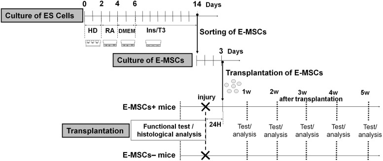
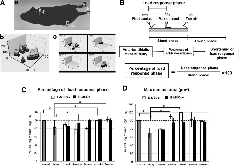
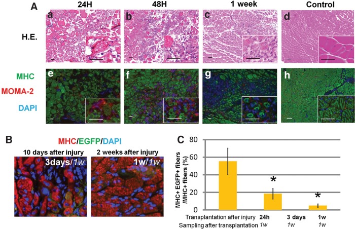
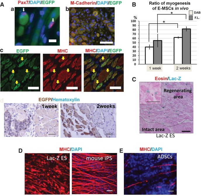
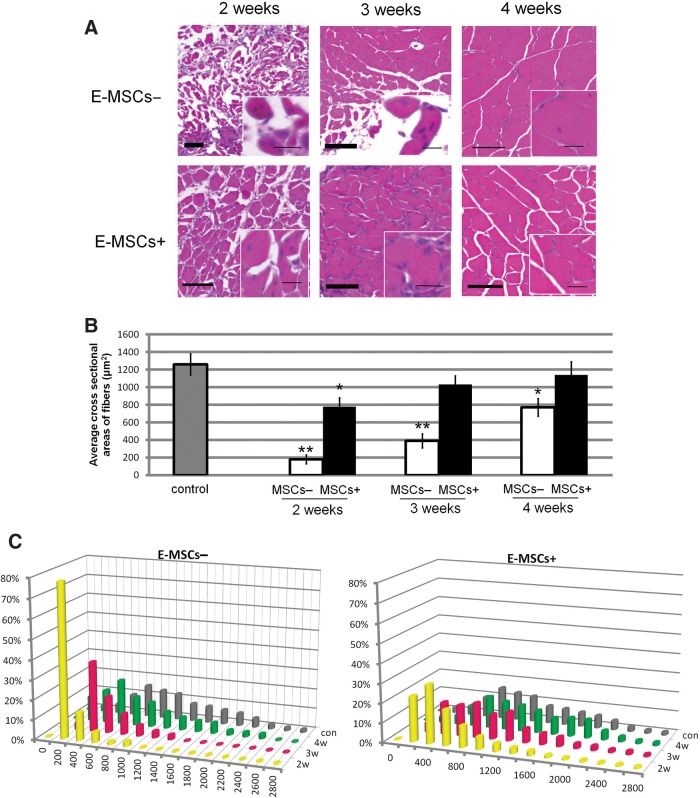
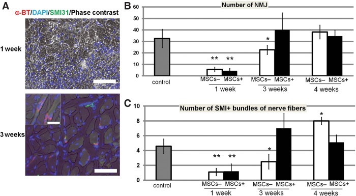
We previously established that mesenchymal stem cells originating from mouse embryonic stem (ES) cells (E-MSCs) showed markedly higher potential for differentiation into skeletal muscles in vitro than common mesenchymal stem cells (MSCs). Further, the E-MSCs exhibited a low risk for teratoma formation. Here we evaluate the potential of E-MSCs for differentiation into skeletal muscles in vivo and reveal the regeneration and functional recovery of injured muscle by transplantation. E-MSCs were transplanted into the tibialis anterior (TA) muscle 24 h following direct clamping. After transplantation, the myogenic differentiation of E-MSCs, TA muscle regeneration, and re-innervation were morphologically analyzed. In addition, footprints and gaits of each leg under spontaneous walking were measured by CatWalk XT, and motor functions of injured TA muscles were precisely analyzed. Results indicate that >60% of transplanted E-MSCs differentiated into skeletal muscles. The cross-sectional area of the injured TA muscles of E-MSC-transplanted animals increased earlier than that of control animals. E-MSCs also promotes re-innervation of the peripheral nerves of injured muscles. Concerning function of the TA muscles, we reveal that transplantation of E-MSCs promotes the recovery of muscles. This is the first report to demonstrate by analysis of spontaneous walking that transplanted cells can accelerate the functional recovery of injured muscles. Taken together, the results show that E-MSCs have a high potential for differentiation into skeletal muscles in vivo as well as in vitro. The transplantation of E-MSCs facilitated the functional recovery of injured muscles. Therefore, E-MSCs are an efficient cell source in transplantation.
我们先前已证实,源自小鼠胚胎干细胞(ES细胞)的间充质干细胞(E-MSCs)在体外分化为骨骼肌的潜力明显高于普通间充质干细胞(MSCs)。此外,E-MSCs形成畸胎瘤的风险较低。在此,我们评估E-MSCs在体内分化为骨骼肌的潜力,并通过移植揭示受损肌肉的再生和功能恢复情况。在直接钳夹后24小时,将E-MSCs移植到胫前肌(TA)中。移植后,对E-MSCs的肌源性分化、TA肌肉再生和再支配进行形态学分析。此外,通过CatWalk XT测量自发行走时每条腿的足迹和步态,并精确分析受损TA肌肉的运动功能。结果表明,超过60%的移植E-MSCs分化为骨骼肌。E-MSC移植动物受损TA肌肉的横截面积比对照动物更早增加。E-MSCs还促进受损肌肉周围神经的再支配。关于TA肌肉的功能,我们发现E-MSCs的移植促进了肌肉的恢复。这是第一份通过对自发行走的分析证明移植细胞可加速受损肌肉功能恢复的报告。综上所述,结果表明E-MSCs在体内和体外均具有分化为骨骼肌的高潜力。E-MSCs的移植促进了受损肌肉的功能恢复。因此,E-MSCs是移植中一种有效的细胞来源。