Park Saeyoung, Choi Yoonyoung, Jung Namhee, Yu Yeonsil, Ryu Kyung-Ha, Kim Han Su, Jo Inho, Choi Byung-Ok, Jung Sung-Chul
Department of Biochemistry, School of Medicine, Ewha Womans University, Seoul 07985, Republic of Korea.
Department of Molecular Medicine, School of Medicine, Ewha Womans University, Seoul 07985, Republic of Korea.
Int J Mol Med. 2016 May;37(5):1209-20. doi: 10.3892/ijmm.2016.2536. Epub 2016 Mar 22.
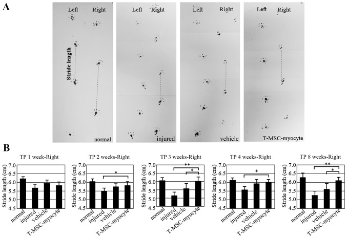
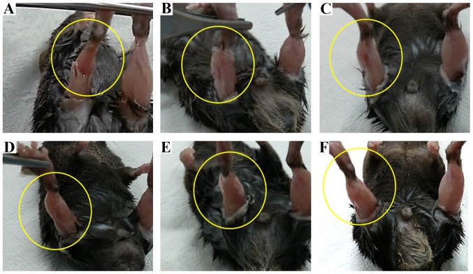
Stem cells are regarded as an important source of cells which may be used to promote the regeneration of skeletal muscle (SKM) which has been damaged due to defects in the organization of muscle tissue caused by congenital diseases, trauma or tumor removal. In particular, mesenchymal stem cells (MSCs), which require less invasive harvesting techniques, represent a valuable source of cells for stem cell therapy. In the present study, we demonstrated that human tonsil-derived MSCs (T-MSCs) may differentiate into myogenic cells in vitro and that the transplantation of myoblasts and myocytes generated from human T-MSCs mediates the recovery of muscle function in vivo. In order to induce myogenic differentiation, the T-MSC-derived spheres were cultured in Dulbecco's modified Eagle's medium/nutrient mixture F-12 (DMEM/F‑12) supplemented with 1 ng/ml transforming growth factor-β, non-essential amino acids and insulin‑transferrin-selenium for 4 days followed by culture in myogenic induction medium [low-glucose DMEM containing 2% fetal bovine serum (FBS) and 10 ng/ml insulin‑like growth factor 1 (IGF1)] for 14 days. The T-MSCs sequentially differentiated into myoblasts and skeletal myocytes, as evidenced by the increased expression of skeletal myogenesis-related markers [including α-actinin, troponin I type 1 (TNNI1) and myogenin] and the formation of myotubes in vitro. The in situ transplantation of T-MSCs into mice with a partial myectomy of the right gastrocnemius muscle enhanced muscle function, as demonstrated by gait assessment (footprint analysis), and restored the shape of SKM without forming teratomas. Thus, T-MSCs may differentiate into myogenic cells and effectively regenerate SKM following injury. These results demonstrate the therapeutic potential of T-MSCs to promote SKM regeneration following injury.
干细胞被视为一种重要的细胞来源,可用于促进因先天性疾病、创伤或肿瘤切除导致肌肉组织结构缺陷而受损的骨骼肌(SKM)再生。特别是间充质干细胞(MSCs),其获取技术侵入性较小,是干细胞治疗中有价值的细胞来源。在本研究中,我们证明了人扁桃体来源的间充质干细胞(T-MSCs)在体外可分化为成肌细胞,并且由人T-MSCs产生的成肌细胞和肌细胞的移植在体内介导了肌肉功能的恢复。为了诱导成肌分化,将T-MSC来源的球体在补充有1 ng/ml转化生长因子-β、非必需氨基酸和胰岛素-转铁蛋白-硒的杜氏改良 Eagle 培养基/营养混合物F-12(DMEM/F-12)中培养4天,然后在成肌诱导培养基[含有2%胎牛血清(FBS)和10 ng/ml胰岛素样生长因子1(IGF1)的低糖DMEM]中培养14天。T-MSCs依次分化为成肌细胞和骨骼肌细胞,体外骨骼肌生成相关标志物[包括α-辅肌动蛋白、肌钙蛋白I 1型(TNNI1)和生肌调节因子]表达增加以及肌管形成证明了这一点。将T-MSCs原位移植到右腓肠肌部分切除的小鼠体内可增强肌肉功能,步态评估(足迹分析)证明了这一点,并且恢复了SKM的形状且未形成畸胎瘤。因此,T-MSCs在损伤后可分化为成肌细胞并有效再生SKM。这些结果证明了T-MSCs在损伤后促进SKM再生的治疗潜力。