Institut Fresnel/UMR-CNRS, D. U. de Saint-Jérôme, Marseille, France.
Comput Math Methods Med. 2013;2013:401413. doi: 10.1155/2013/401413. Epub 2013 Jul 14.
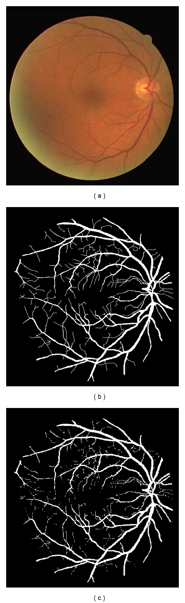
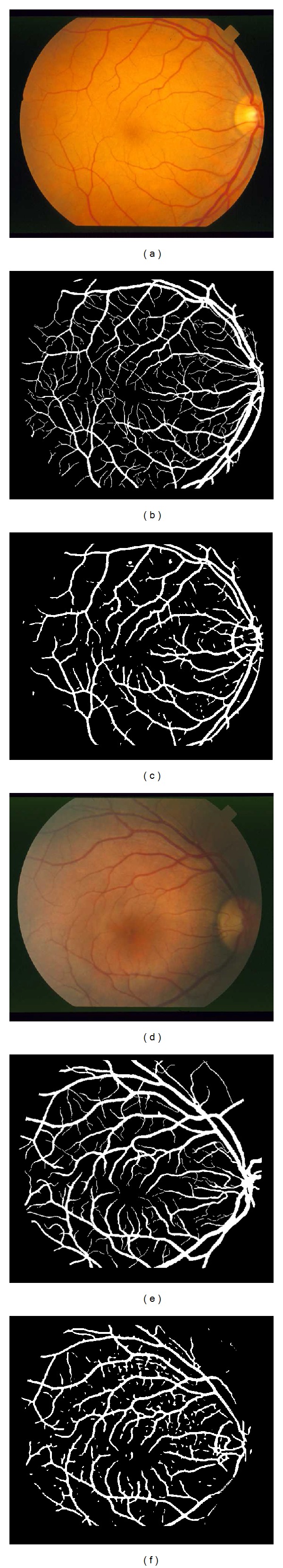
A Bayesian method with spatial constraint is proposed for vessel segmentation in retinal images. The proposed model makes the assumption that the posterior probability of each pixel is dependent on posterior probabilities of their neighboring pixels. An energy function is defined for the proposed model. By applying the modified level set approach to minimize the proposed energy function, we can identify blood vessels in the retinal image. Evaluation of the developed method is done on real retinal images which are from the DRIVE database and the STARE database. The performance is analyzed and compared to other published methods using a number of measures which include accuracy, sensitivity, and specificity. The proposed approach is proved to be effective on these two databases. The average accuracy, sensitivity, and specificity on the DRIVE database are 0.9529, 0.7513, and 0.9792, respectively, and for the STARE database 0.9476, 0.7147, and 0.9735, respectively. The performance is better than that of other vessel segmentation methods.
提出了一种基于贝叶斯方法和空间约束的视网膜图像血管分割方法。该模型假设每个像素的后验概率与其相邻像素的后验概率有关。为该模型定义了一个能量函数。通过应用改进的水平集方法来最小化所提出的能量函数,我们可以识别视网膜图像中的血管。在来自 DRIVE 数据库和 STARE 数据库的真实视网膜图像上对所开发的方法进行了评估。使用包括准确性、敏感性和特异性在内的多种度量标准对性能进行了分析和比较。该方法在这两个数据库上都被证明是有效的。在 DRIVE 数据库上的平均准确性、敏感性和特异性分别为 0.9529、0.7513 和 0.9792,而在 STARE 数据库上的平均准确性、敏感性和特异性分别为 0.9476、0.7147 和 0.9735。该方法的性能优于其他血管分割方法。